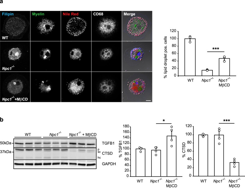
Fig. 7. Lowering cholesterol in Npc1−/− microglia rescues lipid droplet phenotype.
a MβCD treatment (48 h) of cultured Npc1−/− microglia isolated from P7 mice and fed with fluorescently labeled myelin (green) partially restored the formation of lipid droplets as visualized by Nile red (red). Filipin dye (blue) and an anti-CD68 antibody (white) were used to visualize cholesterol and late endosomal/lysosomal compartments, respectively. Scale bar: 10 μm. Cells showing Nile red positive structures at cell periphery were quantified as lipid droplet positive cells. Three independent experiments were analyzed by confocal microscopy (n = 3, p = 0.0003, one-way ANOVA followed by unpaired two-tailed Student’s t-test). b Western blot analysis and quantification of TGFB1 and CTSD (p = pro-form; sc = single chain form; hc = heavy chain form) in cultured Npc1−/− and WT microglia isolated from P7 mice. At 5DIV, Npc1−/− cells were treated with 250 µM MβCD for 48 h. GAPDH was used as a loading control. Protein levels were quantified by densitometry (ImageJ—NIH) from four independent experiments. Values were normalized on WT control and represent mean ± SEM (one-way ANOVA followed by unpaired two-tailed Student’s t-test). TGFB1 (n = 4, p = 0.02); CTSD (n = 4, p = 0.0002).

