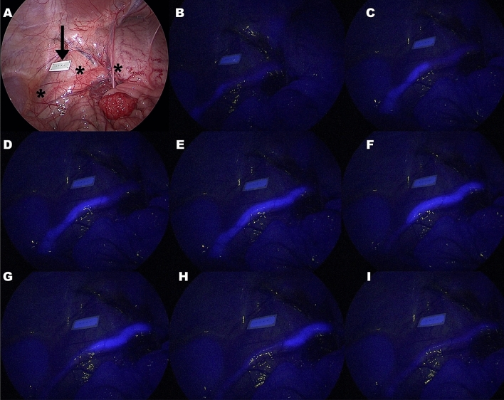
Figure 3.
In (A), demonstration of the IRDye 800BK under white light. The stars indicate the ureter and the arrow the calibration reference card. In a near-infrared mode (B–I), the ureter is not clearly visible before (B) and after (I) a ureteral peristaltic wave (C–H) progresses. The duration of the peristaltic wave is variable, making the fluorescence-aided ureter visualization also variable. In this picture the time between photogram (C) and (H) was approximately of 50 s, whereas photogram (E), the only one showing the ureter entirely, was visible for 3 s, given the progression of the peristaltic wave. For this reason, using the dye the ureter was entirely visible only for 3 s, in this example. Additionally, the high background fluorescence given by the surrounding structures, which de facto limits clear ureter delineation, in absence of the peristaltic wave (B and I) is also visible.

