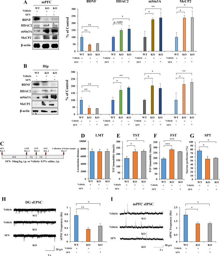
Fig. 4. Nrf2 KO mice show depression-like behavior, alter BDNF, HDAC2, mSin3a, and MeCP2 expression and frequency of sEPSC and sIPSC in hippocampus and mPFC neurons.
A, B The western blot analysis for mPFC and hippocampus. The western blot was performed for Nrf2, BDNF, HDAC2, mSin3A, and MeCP2 in mPFC and hippocampus (mean ± SEM, n = 4 per group, one-way ANOVA, *p < 0.05, **p < 0.01). C The behavior test for Nrf2 KO mice after the SFN treatment. D LMT: locomotion test, E TST: tail-suspension test, F FST: forced swimming test, G SPT: sucrose preference test (mean ± SEM, n = 8 per group, one-way ANOVA, *p < 0.05, **p < 0.01). H Left: representative traces of sEPSC in hippocampal DG neurons. Scale bars represent 2 s, 20 pA. Right: histograms of sEPSC frequency in hippocampal DG neurons (mean ± SEM, n = 10–11 neurons of three mice per group, one-way ANOVA, *p < 0.05, **p < 0.01). I Left: representative traces of sIPSC in mPFC neurons. Scale bars represent 2 s, 50 pA. Right: histograms of sIPSC frequency in mPFC neurons (mean ± SEM, n = 9–10 neurons of three mice per group, one-way ANOVA, *p < 0.05).

