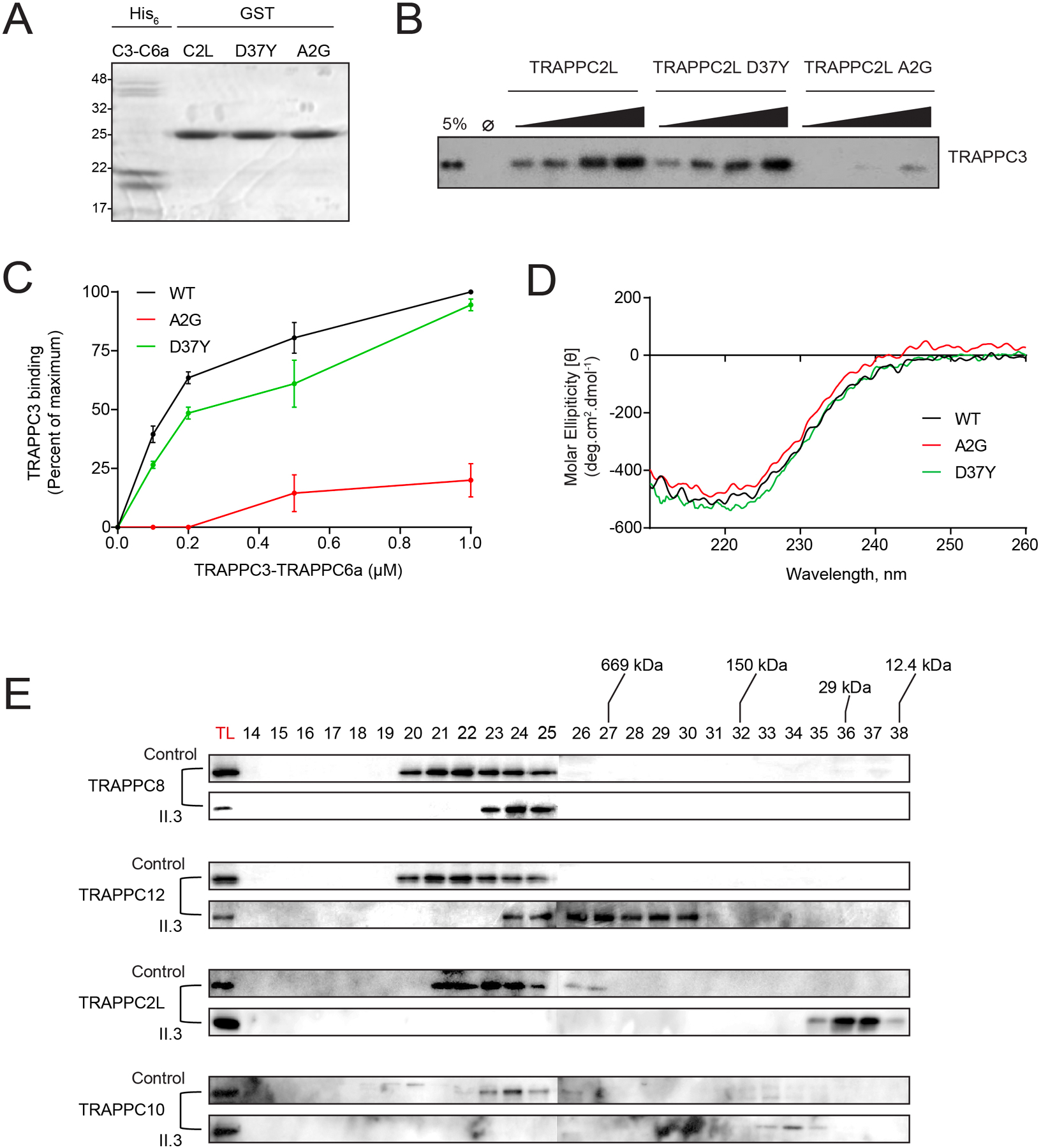
Figure 3. TRAPPC2L p.(Ala2Gly) has a weakened interaction with TRAPPC6a.

(A) an SDS-polyacrylamide gel of His-tagged recombinant TRAPPC3-TRAPPC6a heterodimer and GST-tagged TRAPPC2L or the TRAPPC2L variants p.(Ala2Gly) and p.(Asp37Tyr). Molecular size standards in kD are displayed on the left. (B) The GST-tagged proteins from panel (A) were incubated with increasing concentrations of the TRAPPC3-TRAPPC6a heterodimer. Following binding, the samples were probed by western analysis using anti-TRAPPC3 IgG. A sample representing 5% of the maximum amount of the heterodimer is shown to the left of the panel. (C) The TRAPPC3 signal from panel (B) was quantified and plotted versus the concentration of the heterodimer added to the reaction. (D) A circular dichroism curve for His-tagged recombinant TRAPPC2L or the two variants was performed. (E) Lysates from control fibroblasts (control) and fibroblasts derived from the individual harboring the p.(Ala2Gly) variant (subject) were prepared and subjected to size exclusion chromatography. Total cell lysate (TL) and fractions (indicated above the panels) from the column were analyzed by western analysis for the indicated TRAPP proteins.
