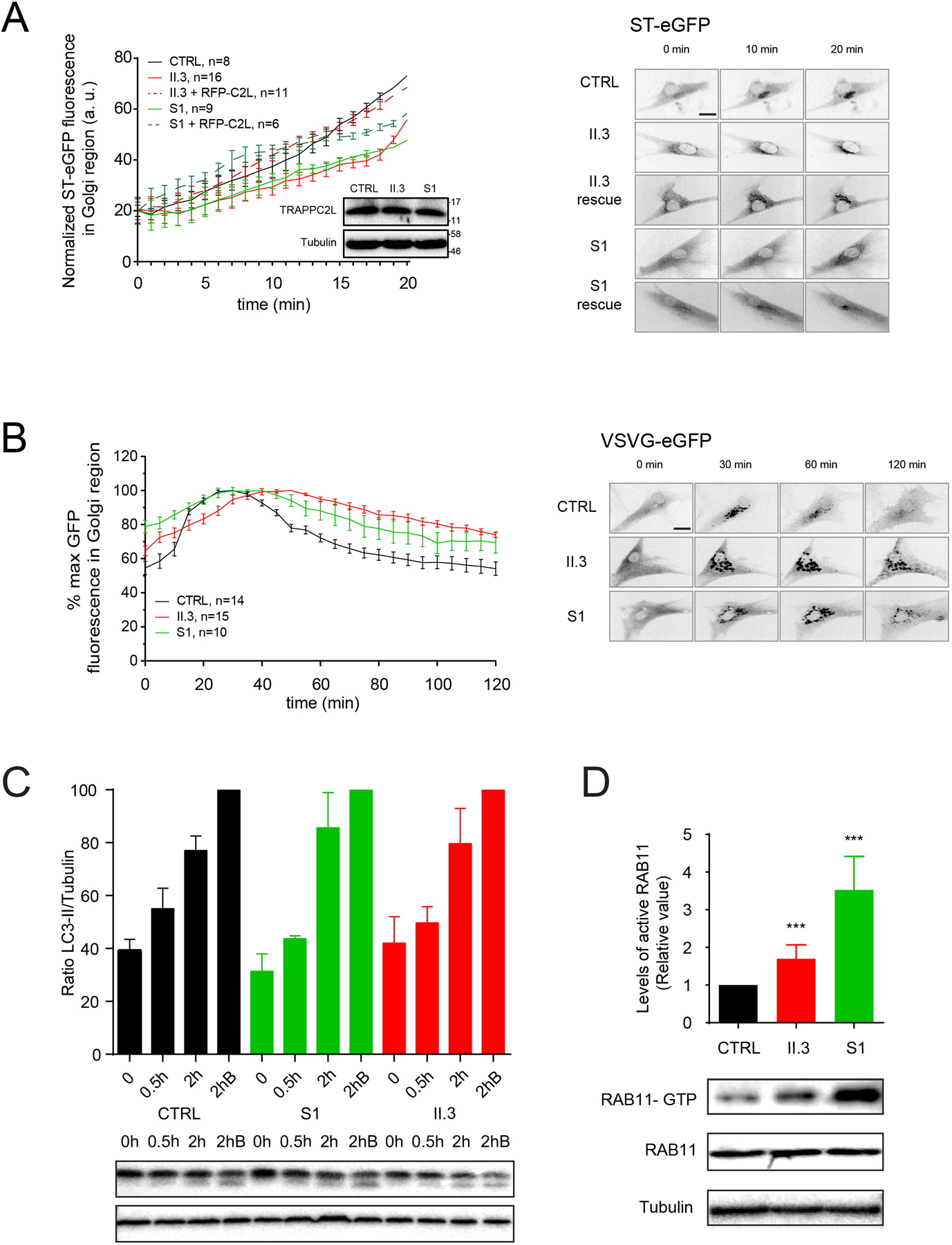
Figure 4. Functional studies reveal a membrane trafficking defect and increased active RAB11 in fibroblasts derived from an individual with TRAPPC2L p.(Ala2Gly).

(A) The RUSH assay measuring traffic between the endoplasmic reticulum and the Golgi was performed and quantified for control (CTRL), p.(Ala2Gly) (II.3) and p.(Asp37Tyr) (S1) fibroblasts using the cargo protein ST-eGFP. The inset shows a western analysis for TRAPPC2L and tubulin from the cells that were analyzed, with molecular size standards on the right. Representative images used for the quantification are shown to the right of the graph. (B) The transport of VSVG-GFP ts045 was performed on control (CTRL), p.(Ala2Gly) (II.3) and p.(Asp37Tyr) (S1) fibroblasts, and quantified. Representative images used for the quantification are shown to the right of the graph. (C) Cells from control (CTRL) and p.(Ala2Gly) (II.3) and p.(Asp37Tyr) (S1) fibroblasts were left in nutrient-rich medium (0) .or starved for 0.5h or 2h. Some cells were starved for 2h in the presence of bafilomycin A1 which prevents formation of autolysosomes (2hB). Lysates were prepared and analyzed by western analysis for the autophagy marker LC3-II as well as tubulin. The normalized LC3-II/tubulin ratio was determined. A sample western blot is shown beneath the graph. (D) Lysates from control (CTRL), p.(Ala2Gly) (II.3) and p.(Asp37Tyr) (S1) fibroblasts were treated with IgG recognizing active (GTP-bound) RAB11. The precipitated RAB11 was then revealed by western analysis using anti-RAB11 IgG. The immunoprecipitated protein is shown I the top panel while the lysates are in the lower two panels. The signal was quantified and plotted as relative levels. *** indicates p<0.001.
