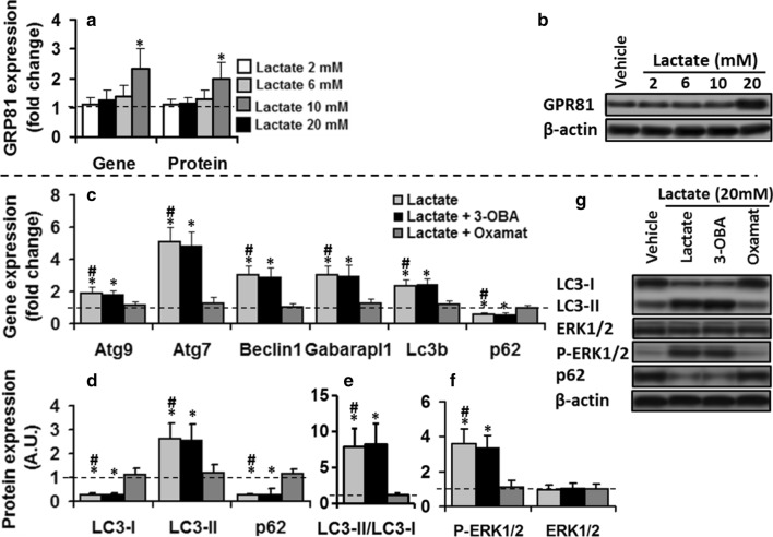
Fig. 5.
Changing the cellular redox associated to lactate oxidation is vital for ERK1/2-mediated autophagic effect of lactate on C2C12 myotubes. C2C12 cells maintained in the vehicle or lactate (2, 6, 10, and 20 mM, 8 h) in the absence or presence of 3-OBA (2 mM) or Oxamate (10 mM) for 8 h. a and b Only 20 mM lactate increased GPR81 gene and protein expression compared to the control values (dashed line). lactate oxidation inhibition by Oxamate abolished lactate stimulatory effect on autophagy-related gene expression c; one-way ANOVA, Atg9, F = 13.5; Atg7, F = 537; Beclin1, F = 34.3, Gabarapl1, F = 23.6; LC3B, F = 29.8, p62, F = 19.4; all p < 0.01), autophagic flux markers (d, e; one-way ANOVA: LC3B-II, F = 23.3; LC3B-II/LC3B-I, F = 142.1; LC3B-I, 32.5; p62, F = 19.4, all p < 0.01), and P-ERK 1/2 (f), inhibition of GPR81 by 3-OBA had no effect. The values expressed as mean ± SD of fold change from basal values (dashed line). For all protein measurement, fold changes in protein levels were normalized for β-actin and relative to their expression in basal condition (dashed line). * Significant difference with basal values (P < 0.01), # Significant difference with lactate + oxamate group (P < 0.01), N = 6 per group

