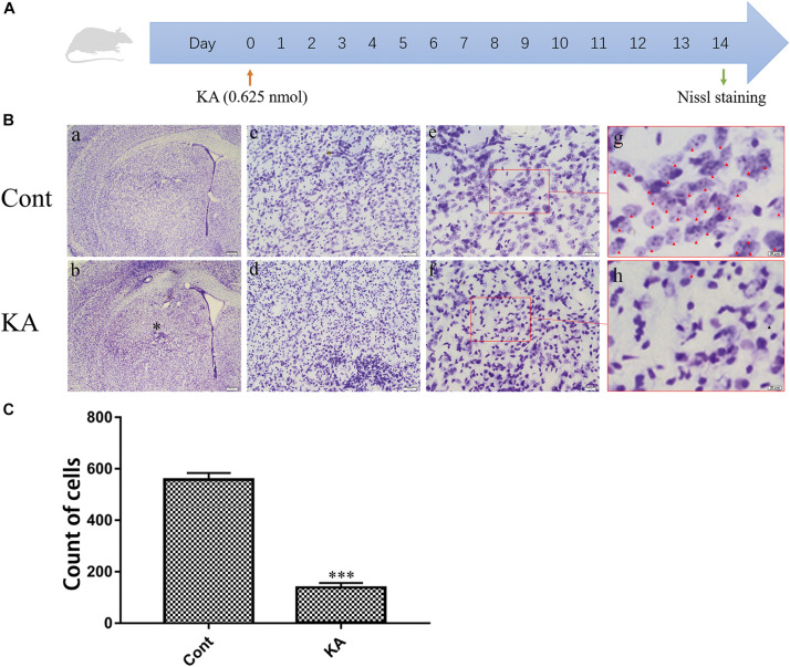
FIGURE 1.
KA causes neuronal injury in the striatum. (A) Illustration of experimental design. Mice were sacrificed on day 14 after KA injection. Nissl staining were performed on brain sections. (B) Representative microscopic images of Nissl-stained coronal brain section displaying the lesion. (C) Quantification of neurons with normal morphological specificity. The histological analyses were single-blinded. Data are expressed as the Mean ± SEM. Scale bar = 500 μm in a, b; 100 μm in c, d; 50 μm in e, f; and 20 μm in g, h (n = 5. ***P < 0.001 vs control, in Student’s t test).

