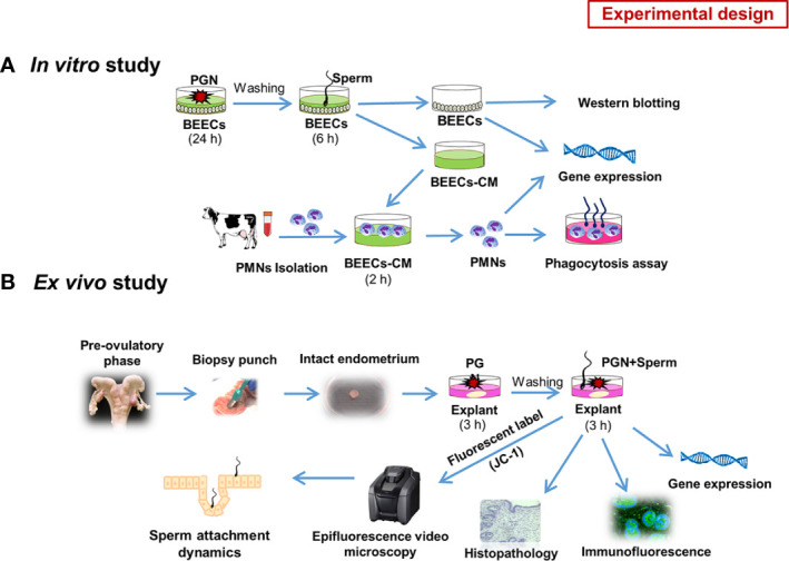
Figure 1.

Schematic representation of the study experimental design: (A) in vitro model. Initially we investigated the competitive interaction of LPS/PGN and sperm with endometrial epithelium using RT-PCR. Based on the results, we gave more focus on the impact of low concentrations of PGN on sperm-induced inflammation in endometrial epithelium. As a main experiment, Subconfluent BEEC monolayers (90%) were stimulated with PGN (10 pg ml−1) for 24 h, followed by co-culture with sperm (5 × 10⁶ ml−1) for 6 h. The impact of PGN on sperm-induced immune responses in BEECs was assessed using RT-PCR. Later on, sperm-triggered MAPK cascade components in BEECs were assessed using western blotting. Conditioned medium (CM) from a sperm-BEEC co-culture after pre-exposure to PGN was harvested and exploited to challenge peripheral PMNs, isolated from mature healthy cows, for 2 h. Subsequently, PMN immune responses and phagocytic activity toward sperm were measured (A). To assess direct BEEC response to PGN alone, BEECs were exposed to different concentrations of PGN at different time points. For activation or blockage of the TLR2 pathway, a TLR2 agonist (pam3Cys) or antagonist (CU-CPT22) was used, respectively. (B) ex vivo model. An explant model of intact endometrium was used to explore the sites of interactions and sperm dynamics with endometrial epithelium. We investigated immune responses and histomorphology induced by sperm in endometrial tissue in the presence of PGN. Additionally, we used high-resolution 3D imaging microscopy to trace sperm dynamics and attachment after JC-1 mitochondrial illumination of their mid-pieces (B).
