Figure 8.
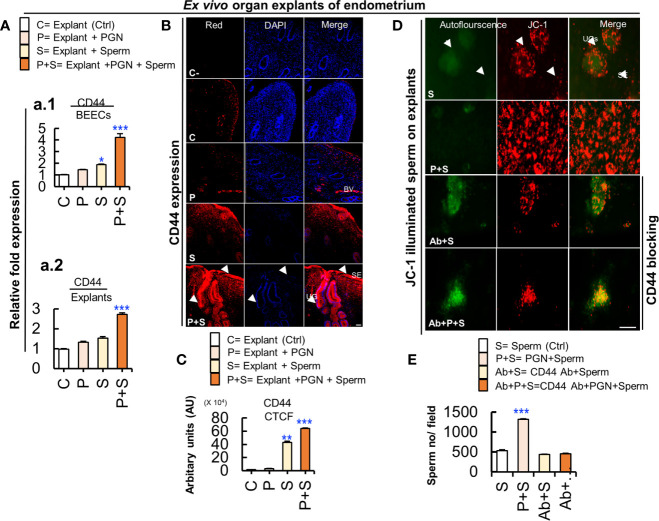
Cluster of differentiation 44 (CD44) is expressed in the bovine endometrium and is implicated in the massive PGN-triggered sperm attachment to endometrial glands and luminal epithelium. (A) RT-PCR assay revealed upregulated CD44 mRNA expression in BEECs in vitro (a.1) and in endometrial tissue ex vivo (a.2). Data are presented as mean ± SEM of 3 independent experiments using epithelial cells or explants from 3 different uteri (3 wells/treatment/experiment). Asterisks denote a significant variance (*P < 0.05, or ***P < 0.001). (B) Immunofluorescence assay of cellular adhesion molecule (CD44) in ex vivo organ explants of endometrium. Immunostaining confirmed intense accumulation of CD44 (red) (arrow heads) induced by PGN+Sperm (“P+S” group) specifically in UGs and in SE (arrow heads) compared to sperm “S” and other groups (C and P). Magnification: 200×. Scale bar = 50 μm. (C) Semiquantitative scoring of corrected total cell fluorescence (CTCF) of CD44 expressed in UGs as arbitrary fluorescence units (AU). Data are presented as mean ± SEM of 3 independent experiments using explants from 3 different uteri (3 wells/treatment/experiment). Asterisks denote a significant variance (**P < 0.01 or ***P < 0.001). (D) PGN triggered massive sperm association to uterine glands (UGs) (arrows) and surface epithelium (SE) (arrow heads) compared to control (sperm). Pre-exposure of endometrial explants to anti-rabbit CD44 polyclonal antibody for 3 h followed by exposure to PGN (10 pg ml−1) for 3 h, and co-culturing with sperm for 3h (Ab+P+S) resulted in minimizing PGN-triggered sperm attachment (P+S) in endometrial explants and most of spermatozoa were released actively and found freely swimming on the surface epithelium (Ab+PGN+S) ( Supplementary Video 4 ). Magnification: 200×. Scale bar = 200 μm. (E) ImageJ software (version 1.51j8) was used for counting localized spermatozoa in ( Supplementary Videos 1 and 2 ). Data are presented as mean ± SEM of 3 independent experiments using explants from 3 different uteri. Asterisks denote a significant variance (***P < 0.001) between the different groups compared to control (sperm only).

