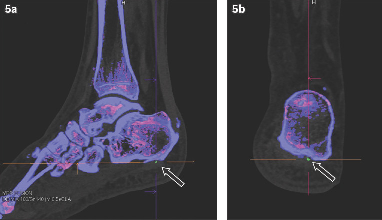
Fig. 5.

A 67-year-old man with a history of hyperuricaemia presented with chronic intermittent left heel pain. (a) Sagittal and (b) coronal post-processed dual-energy CT images show monosodium urate deposition, depicted in green, in the plantar fascia (arrows).
