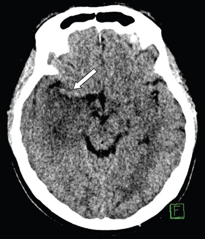
Fig. 14.

Non-contrast CT image of the brain shows a focal area of hypodensity at the amygdala and hippocampus of the right temporal lobe, which is due to limbic encephalitis. The right middle cerebral artery gives a false impression of hyperdensity relative to the adjacent hypodense right temporal lobe (arrow).
