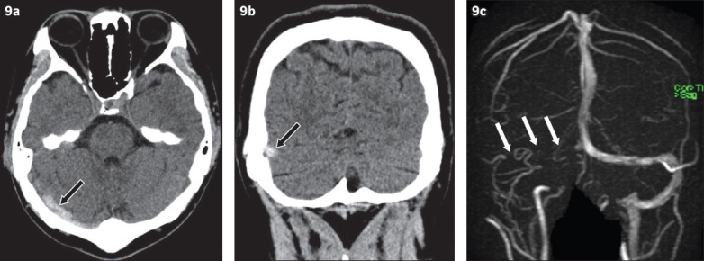
Fig. 9.

Non-contrast CT images of the brain taken in the (a) axial and (b) coronal planes show the hyperdense vessel sign in the right transverse sinus (arrow in a & b) of a patient who presented with severe headache. (c) Phase contrast MR venography maximum intensity projection image confirms the finding of transverse sinus thrombosis by demonstrating an absence of flow signal in the expected region of the right transverse sinus (arrows).
