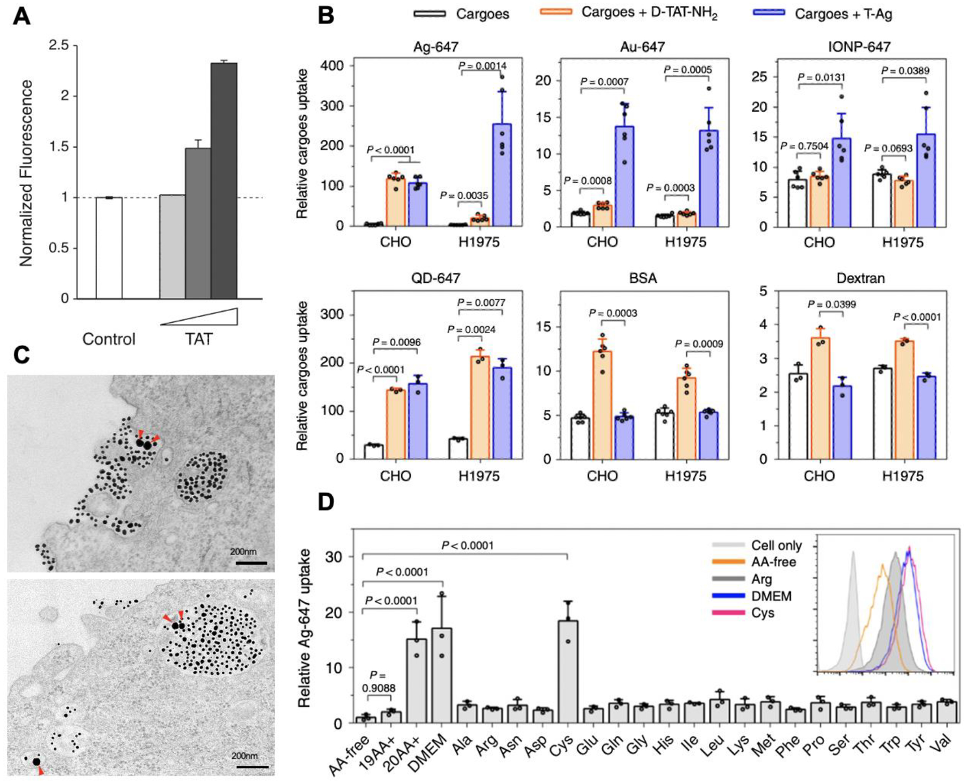
Figure 3. Receptor-mediated macropinocytosis and bystander uptake.

(A) TAT peptide stimulated the bystander uptake of Dextran. The image is adapted from [73].
(B) The bystander uptake of the indicated bystander cargo by TAT peptide (orange bars) and TAT-NPs (blue bars). The image is adapted from [8].
(C) TEM images shows that TAT-coated gold NPs (smaller dark dots) and bystander gold NPs (larger dark dots, pointed by red arrows) are engulfed together by macropinocytosis-like vesicles. Scale bar = 200 nm. The image is adapted from [8].
(D) Extracellular cysteine stimulated TAT-NP-induced bystander uptake. Cells were incubated with TAT-NPs and bystander AgNPs in the culture media containing various amino acid compositions: no amino acid (AA-free), the indicated amino acid alone (e.g. Cys, Arg), 19 amino acids without cysteine (19AA+) and all the amino acids (20AA+). TAT-NP-mediated bystander uptake is much higher in the media containing all 20 AAs or only cysteine, than that in the media without Cysteine. The image is adapted from [8].
