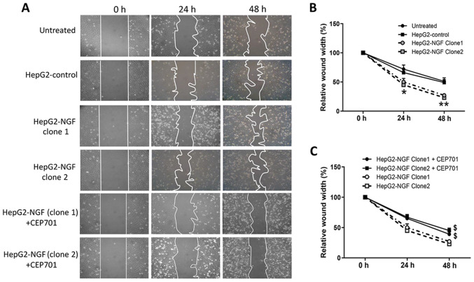
Figure 2.
Effect of NGF on cell motility. Wound healing assay were used to examine the cell motility. (A) Images were acquired at 0, 24 and 48 h using an inverted microscope (10× magnification) after the confluent cells were wounded by scratching cell sheets with a 10 µl pipette tip. CEP701 was added at 0 h. (B) The wound closure was quantitatively analyzed using ImageJ software by outlining and assessing the unhealed area in the wound images. Data are presented as the mean ± SEM, n=4. *P<0.05, **P<0.01 vs. HepG2-control and untreated group (one-way ANOVA). (C) Effect of CEP701 on cell motility. Data are presented as the mean ± SEM, n=4. $P<0.05 vs. HepG2-NGF cells without CEP701 treatment (one-way ANOVA). NGF, nerve growth factor.

