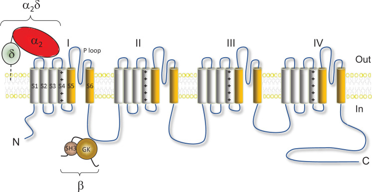
Figure 1. Schematic representation of the structure of CaV channels.
The CaVα1 subunit is formed by four repeat domains (I–IV) each containing six transmembrane segments: S1–S4 constitute the voltage sensor domain (S4 segments contain positively charged residues) and S5–S6 constitute the pore domain (the P loops contain acidic residues that contribute to the selectivity filter of the channel). CaVα1 subunits can be associated with auxiliary subunits: an extracellular α2δ subunit attached to the plasma membrane by a glycosyl phosphatidylinositol (GPI) anchor and an intracellular β subunit which contain a src homology 3 (SH3) domain and a GK domain.

