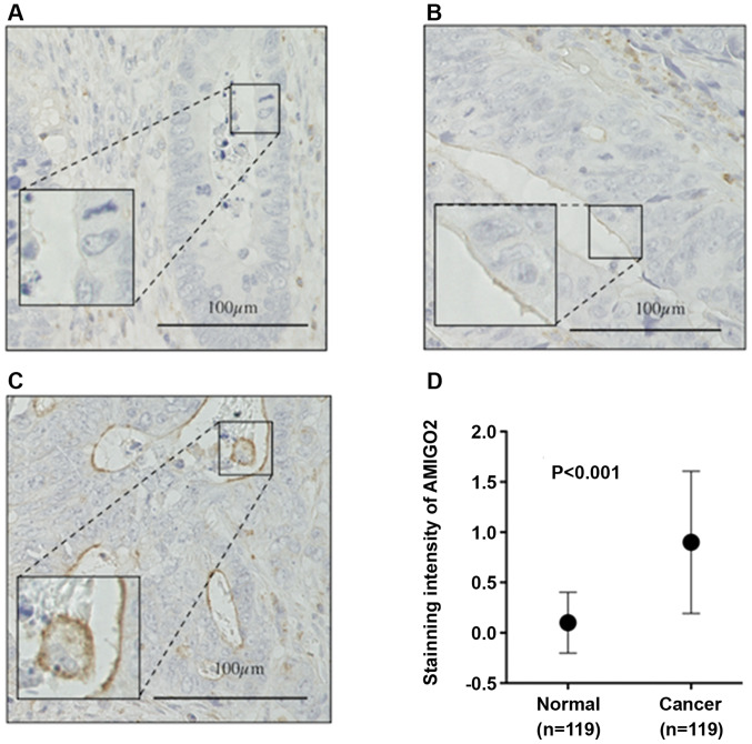
Figure 3.
Immunohistochemistry. Representative pictures of AMIGO2 cell surface staining in colorectal cancer cells for the different intensity scores used in this study. (A) Score 0 (negative), (B) score 1 (weak), and (C) score 2 (moderate to strong). (D) The intensity of AMIGO2 staining was significantly stronger in cancer tissue, when compared with normal tissue (P<0.001). The Wilcoxon test was used for the statistical analysis. AMIGO2, adhesion molecule with Ig like domain 2.

