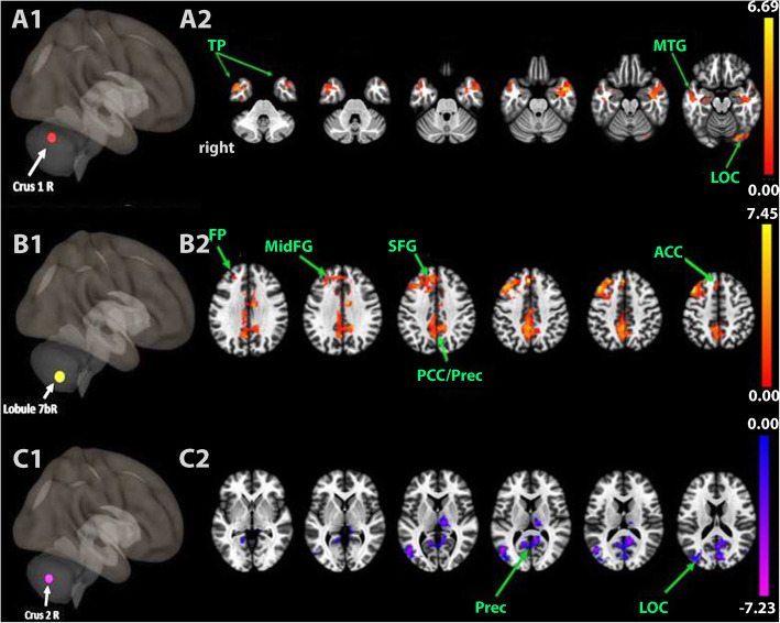
Fig. 1.
Static resting-state functional connectivity map between seeded right lobule VII and nodes of intrinsic networks (p-FDR < 0.05, cluster-level) after real tDCS stimulation compared with sham tDCS. A1-C1. Schematic lateral 3D-view showing the seeded cerebellar lobules. A2-C2. Axial slices passing through the brain. A2. Increased functional connectivity between crus 1 and visual/temporal regions. B2. Increased functional connectivity between lobule 7b and nodes belonging to SN (ACC), DMN (PCC, Precuneus) and CEN (FP, MidFG). C2. decreased functional connectivity between crus 2 and visual/ DMN regions. The colored bars represent the z-value

