Fig. 6.

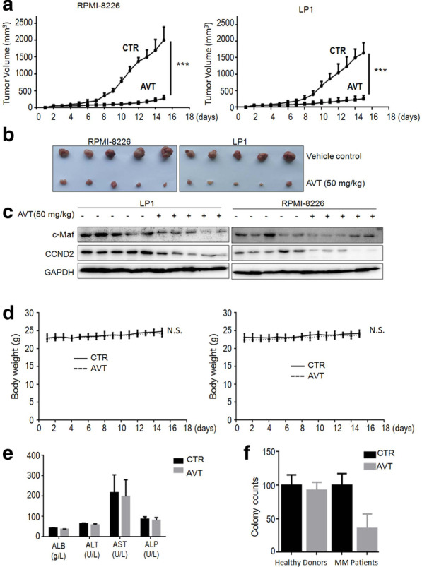
AVT delays myeloma tumor growth in immunodeficiency mice. Two myeloma xenograft models were established by injection of RPMI-8226 or LP1 cells into immunodeficiency mice. a Tumor volume growth was monitored every day during 15 d of treatment. Data were expressed as mean ± standard deviation. ***; p < 0.001. b At the end of the experiment, tumor tissues were dissected. c Tumor tissues from each model were subjected to WB analyses for c-Maf, CCND2, and GAPDH with specific antibodies. d Mice bodyweight was measured every day during AVT treatment. e albumin (ALB), alanine amino transferase (ALT), aspartate amino transferase (AST), and alkaline phosphatase (ALP) from mice blood were analyzed at the end of the experiment. f Mononuclear cells from bone marrow collected from healthy donors or MM patients were treated with AVT before being plated in MethoCult® GF H4434 medium to allow colony formation. Colonies were counted on the 14th day of treatment. Colonies with more than 20 cells were counted for analysis. CTR, vehicle control; AVT, acevaltrate
