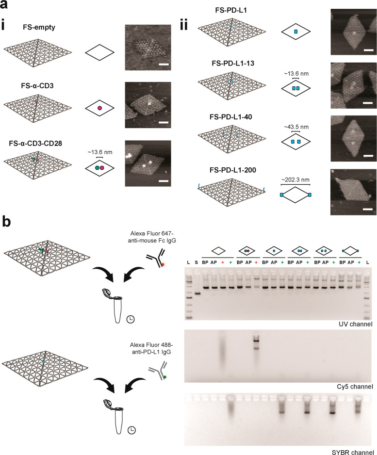
Figure 2.
Production of protein–DNA flat sheets. (a) (i) Schematic designs of DNA flat sheets without proteins (FS-empty), functionalized with one anti-CD3 IgG–oligo conjugate in the center (FS-α-CD3), anti-CD3 IgG– and anti-CD28 IgG–oligo conjugates (FS-α-CD3-CD28), and (ii) functionalized with PD-L1–oligo conjugates at different positions (FS-PD-L1, FS-PD-L1-13, FS-PD-L1-40 and FS-PD-L1-200). For simplistic representation, flat sheets are depicted as rhombi and anti-CD3 IgG, anti-CD28 IgG, and PD-L1 are shown as magenta, green, and cyan blobs, respectively. Representative AFM images of flat sheets folded in 1× PBS (right column). Scale bar = 50 nm. (b) Immunolabeling of protein–DNA flat sheets with fluorescently labeled antibodies and agarose gel electrophoresis. L, 1 kb Plus DNA ladder. S, p8064 ssDNA scaffold. BP, before Sepharose purification. AP, after Sepharose purification. Red plus symbol, addition of Alexa Fluor 647-anti-mouse Fc IgG to flat sheets. Green plus symbol, addition of Alexa Fluor 488-anti-PD-L1 IgG to flat sheets.

