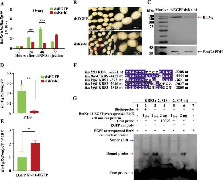
Fig. 2.
Effects of RNAi mediated silencing of BmKr-h1 on the BmVgR expression and the oocyte development. A total of 10 μg of dsRNA per pupae was injected into the intersegmental region in the abdomen of 6-day-old pupae. A qRT-PCR analysis of BmKr-h1 mRNA in the ovaries of the treated pupae. B Phenotypes of oocytes in the ovaries of the dsRNA-treated pupae at day 8. C Western blot analysis of BmVg protein in the oocyte at 48 h post dsRNA treatment. A total of 15 μg protein was loaded per lane and probed with anti-BmVg and anti-GAPDH antibodies, respectively. D qRT-PCR analysis of the expression change of BmVgR in the oocyte. E qRT-PCR detection of the effect of BmKr-h1 overexpression on the transcript level of BmVgR. F Prediction of KBS in the BmVgR promoter based on the KBS core sequences of the BmBR-C and BmE93 promoters [3, 31]. The conserved amino acid residues are in blue background. G EMSA of the binding of the nuclear protein from BmN cells overexpressing BmKr-h1-EGFP with the KBS3 in the BmVgR promoter. For qRT-PCR, Rp49 amplified was used as the internal control. Significance of the differences between the treatment and control was statistically analyzed at p < 0.05 (*), p < 0.01 (**), and p < 0.001 (***) using t test. Dn, day of development; P, pupal stage

