
Fig. 2 |. Structure of apo-FPN.
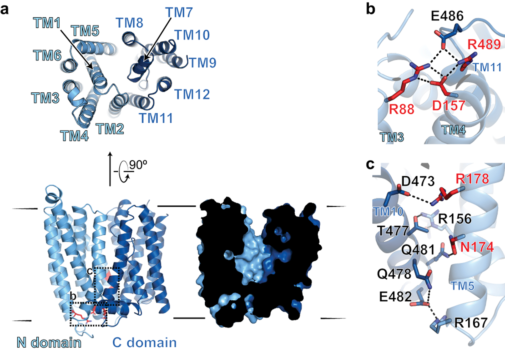
a, Ribbon diagram of FPN reveals 12 transmembrane helices. The N- and C-domains are colored in different shades of blue. Cutaway surface view (right) shows outward open conformation. b, Intracellular gating residues are shown as sticks. c, TM10 and TM5 form an extensive network of interactions, further stabilizing the outward open conformation. Residues highlighted in red in (b) and (c) are known loss-of-function mutations that lead to ferroportin disease in humans.