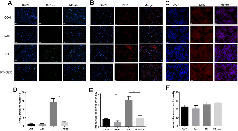
Figure 3.
Effect of DZR on post-radiation ROS generation in heart and tumor tissue. (A, D) show typical TUNEL-stained photomicrographs of heart tissues (×400, n=7–11 respectively). TUNEL- and DAPI-positive cells appear green and blue, respectively. Myocardial cell apoptosis increased markedly in RT rats compared to other groups. (B) Representative images of dihydroethidium (DHE) fluorescence staining (×400) of rat cardiomyocytes. DHE=red, nuclei=blue. (E) Quantitative analysis of ROS (n=5 per group) in rat heart tissues. (C) Representative microphotographs of DHE staining (×400) in tumor tissues from nude mice. (F) No differences in DHE fluorescence density of tumor tissues were observed in quantitative analysis (n=5 per group). mean ± SEM, *: p <0.05, **: p <0.01. scale bar =50 μm.

