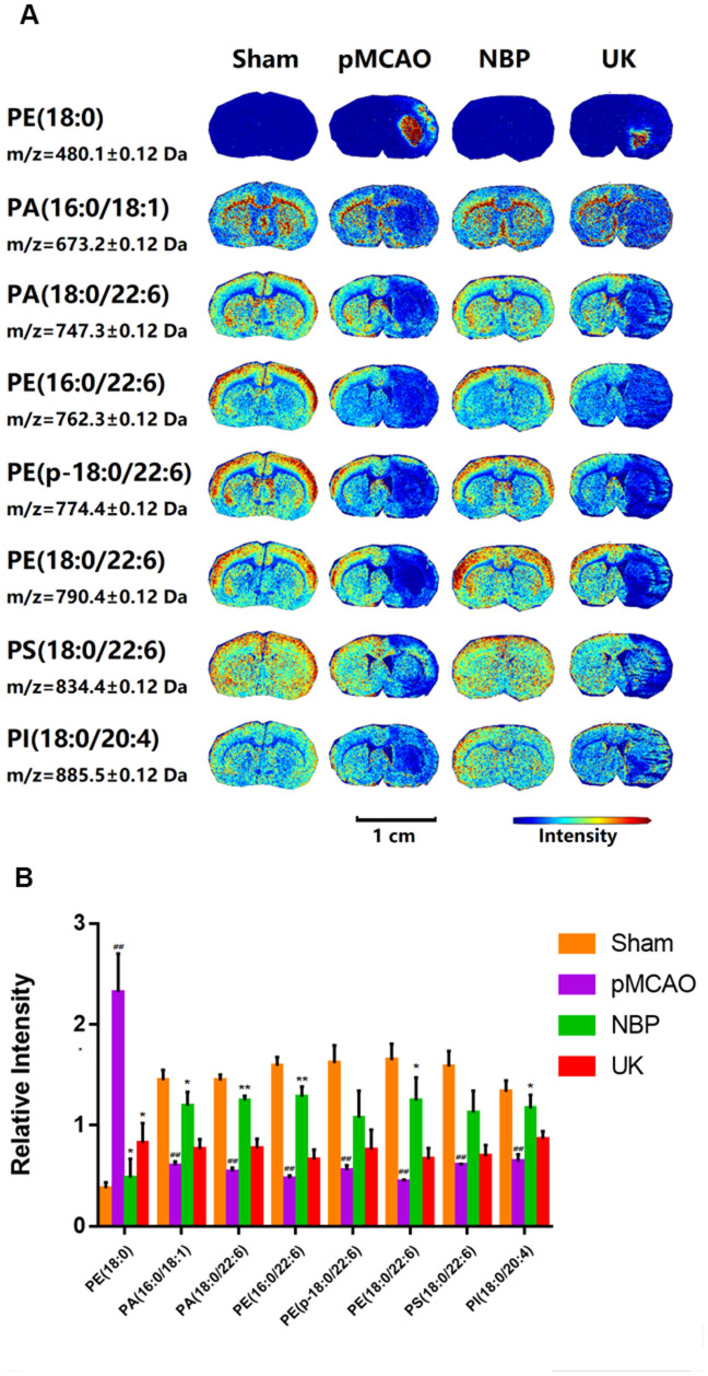
Figure 2.
Changes in phospholipid levels in the brains of rats with pMCAO. (A) In situ MALDI-TOF MSI of phosphatidylethanolamine (PE) (18:0), phosphatidic acid (PA) (16:0/18:1), PA (18:0/22:6), PE (16:0/22:6), PE (p-18:0/22:6), PE (18:0/22:6), phosphatidylserine (PS) (18:0/22:6) and phosphatidylinositol (PI) (18:0/20:4). The spatial resolution was set to 100 μm. Scale bar = 1 cm. (B) Statistical analysis of the relative intensities of the phospholipids mentioned above. Values were normalized to those in the left brain where there was no ischemia. The data are presented as the mean ± SD, n = 3, and were assessed using one-way ANOVA. ## P < 0.01 vs. the sham group, * P < 0.05 vs. the pMCAO group, ** P < 0.01 vs. the pMCAO group.

