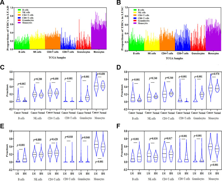
Figure 6.
Correlations between TSKU methylation and the proportions of infiltrating immune cells in LUAD and LUSC. (A) The proportions of tumor-infiltrating immune cells (TIICs) in every sample using the TCGA Infinium 450K methylation data in LUAD (N = 460). (B) The proportions of TIICs in every sample using the TCGA Infinium 450K methylation data in LUSC (N = 372). (C) Comparing the proportions of TIICs in tumor tissues (N = 460) and normal tissues (N = 32) in LUAD datasets. (D) Comparing the proportions of TIICs in tumor tissues (N = 372) and normal tissue (N = 43) in LUSC datasets. (E) Comparing the proportions of different TIICs between groups with high TSKU methylation levels (N = 230) and low TSKU methylation levels (N = 230) in LUAD samples. (F) Comparing the proportions of different TIICs between groups with high TSKU methylation levels (N = 185) and low TSKU methylation levels (N = 185) in LUSC samples (LM, low methylation; HM, high methylation).

