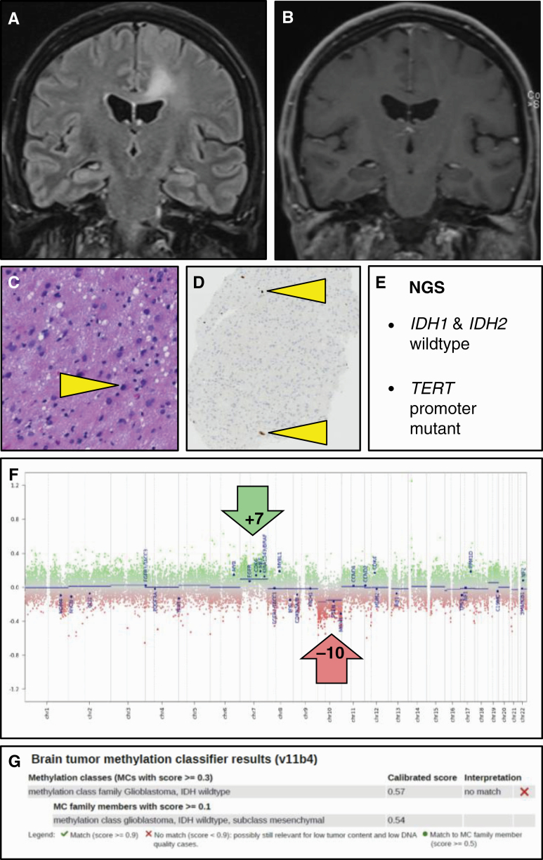
Figure 3.
Histologically low-grade isocitrate dehydrogenase (IDH)–wild-type astrocytoma with molecular features of glioblastoma. A, T2–fluid-attenuated inversion recovery MRI of this 54-year-old male patient reveals a lesion that is compatible with diffuse low-grade glioma; in B, T1-weighed postgadolinium scans, the lesion is not enhancing. C and D, Hematoxylin-eosin staining of the needle biopsy material reveals only a slight increase in cellularity with mild nuclear pleomorphism and variably increased size of the (eosinophilic) cytoplasm, while Ki-67 staining reveals only very few proliferating cells (some positive nuclei indicated by arrowheads); these histopathological findings are compatible with the diagnosis of diffuse low-grade glioma. E, Next-generation sequencing of this lesion, however, reveals the absence of IDH1 and IDH2 mutation, but the presence of a TERT promoter mutation, meaning that the lesion according to cIMPACT-NOW update 3 qualifies as glioblastoma, IDH–wild-type, F, The fact that the copy number profile (obtained by performing methylome analysis) reveals gain of complete chromosome 7 combined with loss of complete chromosome 10 fully supports this diagnosis. G, Analysis of the methylome profile of this lesion using the Heidelberg Brain Tumor Classifier14,15 also suggests the diagnosis glioblastoma, IDH–wild-type as best match; the low tumor cell content in this biopsy material may well explain the suboptimal score for this diagnosis (as well as the relatively subtle levels of chromosome 7 gain and chromosome 10 loss in F). cIMPACT-NOW, Consortium to Inform Molecular and Practical Approaches to CNS Tumor Taxonomy—Not Official WHO.

