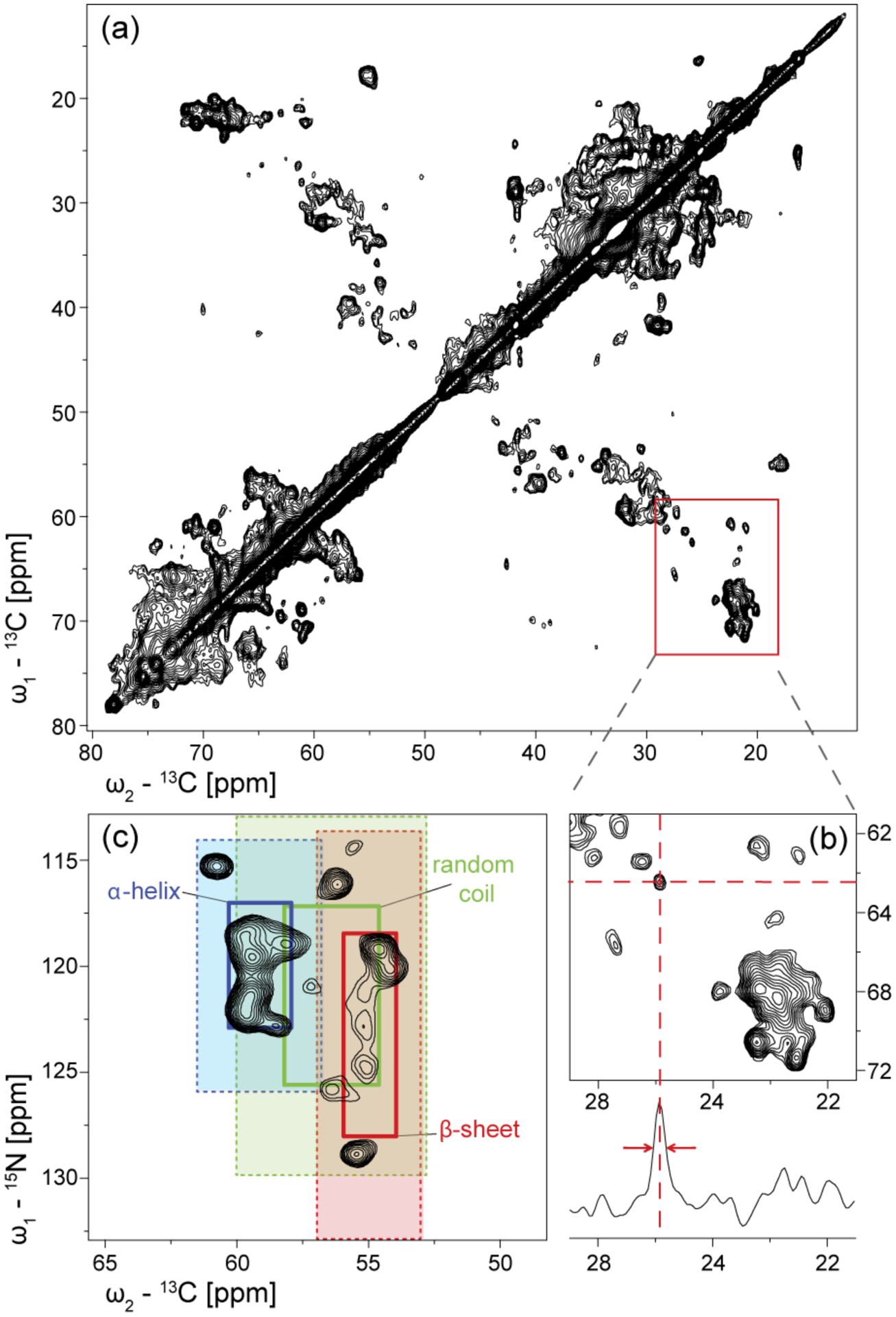
Figure 5.

(a) Aliphatic region of the 13C-13C PDSD spectrum (20 ms mixing time) of [13C,15N-DEQGHKTS]-MsbA reconstituted in DMPC/DMPA (9:1) at LPR=50 mol/mol. Labelling was achieved using extensive reverse labelling (see materials and methods). The homogeneous sample preparation results in a number of individually resolved cross peaks. (b) A 1D trace taken at 62.5 ppm shows a 0.5 ppm linewidth (106 Hz) at half height for an isolated peak. (c) 15N-13C NCA spectrum of [13C, 15N-K]-MsbA with secondary structure regions highlighted (1- and 2-times standard deviation) (Wang & Jardetzky, 2002). MsbA contains 22 lysines.
