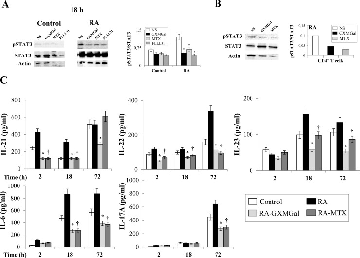
Fig 6. GXMGal effect on Th17 response.
Activated PBMC (A and C) or purified CD4+ T cells (B) (both 5×10⁶/ml) from Control and RA were incubated for 2, 18 and 72 h in the presence or absence (NS) of GXMGal (10 μg/ml), MTX (10 ng/ml) or FLLL31 (5 μM). After 18 h of incubation, cell lysates were analyzed by western blotting. Membranes were incubated with Abs to pSTAT3 and STAT3. Actin was used as an internal loading control. Normalization was shown as mean ± SEM of five independent experiments (A) or as one representative experiment of three with similar results (B). *, p<0.05 (triplicate samples of 5 different Control and RA; RA treated vs untreated cells). Note that for both Control and RA cells in (A), the immunoblots share Actin loading control panels with the experiments shown in Fig 1A at 18 h, as the same immunoblots were stripped and re-probed using different antibodies. Culture supernatants were collected after 2, 18 and 72 h to test IL-21, IL-22, IL-23, IL-6 and IL-17A levels by specific ELISA assays. *, p<0.05 (triplicate samples of 7 different Control and RA; RA GXMGal-treated vs untreated cells); †, p<0.05 (triplicate samples of 7 different Control and RA; RA MTX-treated vs untreated cells) (C).

