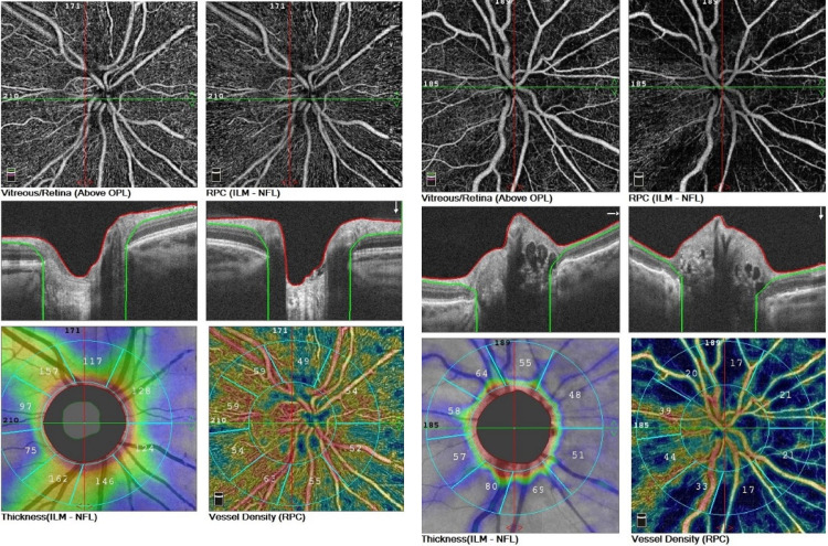
Fig 3. The optic disc OCT-A of the right eyes, in eye with optic disc drusen (the left side) and the control group (the right side).
In sequence: nonperfusion areas in the radial peripapillary capillaries; B-scans of optic nerve head; coloured coded density maps; reduced RNFL and vascular density consistent with the blue areas.

