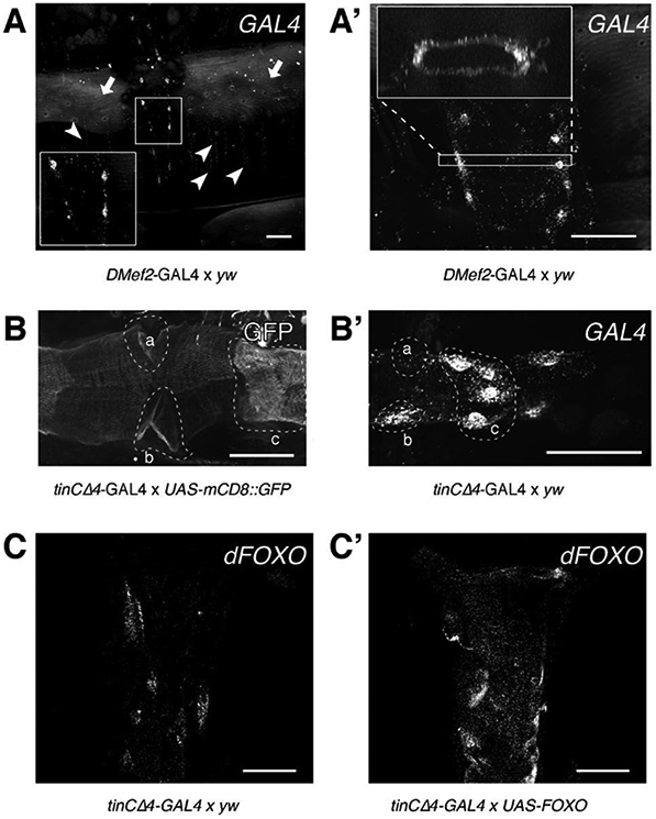
Fig. 2. Characterization of GAL4 expression patterns in DMef2-GAL4 and tinCΔ4-Gal4 using RNAscope and ViewRNA.

(A) Adult Drosophila abdominal carcass treated with a probe against GAL4 shows expression of GAL4 in abdominal body wall muscles (arrowheads) and in the cardiomyocytes of the heart (inset). Note the background fluorescence from the cuticle (arrows). (A’) RNA molecules can be detected throughout the heart tube (cross-sectional view). (B) Activity of GAL4 assayed via mCD8::GFP reporter gene expression. GFP can be detected throughout the entire heart tube, but shows stronger fluorescence in the ostia cells (a, b) and valve cells (c). (B’) GFP intensity is mirrored by high levels of GAL4 transcripts in the same cell types (a, b, and c, compare to B). (C) Endogenous dFOXO transcripts from control hearts (tinCΔ4-GAL4 × yw) were probed using ViewRNA. (C’) tinCΔ4-GAL4 drives UAS-dFOXO expression to a visibly greater extent than the endogenous dFOXO promoter in control hearts. All scale bars (white) represent 50 μm. (C and C’) were adapted for use with permission from [23].
