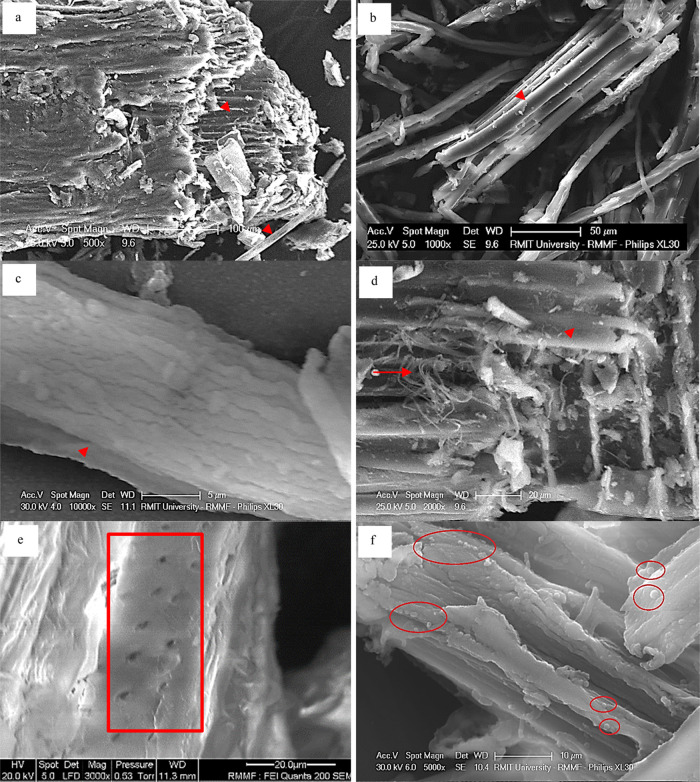
Figure 7.
Distinguishing features of Eucalyptus sawdust showing intact macrofibrils within fibers (a); exposed macrofibrils of 5–20 μm width (b); exposed macrofibrils encasing microfibrils (c); exposed microfibrils of 0.1–2.0 μm width (d); with surface micropores of 1–5 μm (e); and lignin nanospheres of 0.5–2.0 μm (f). Macrofibrils are marked by arrowheads, microfibrils marked by arrows, surface micropores marked by a box, and nanospheres marked by circles.

