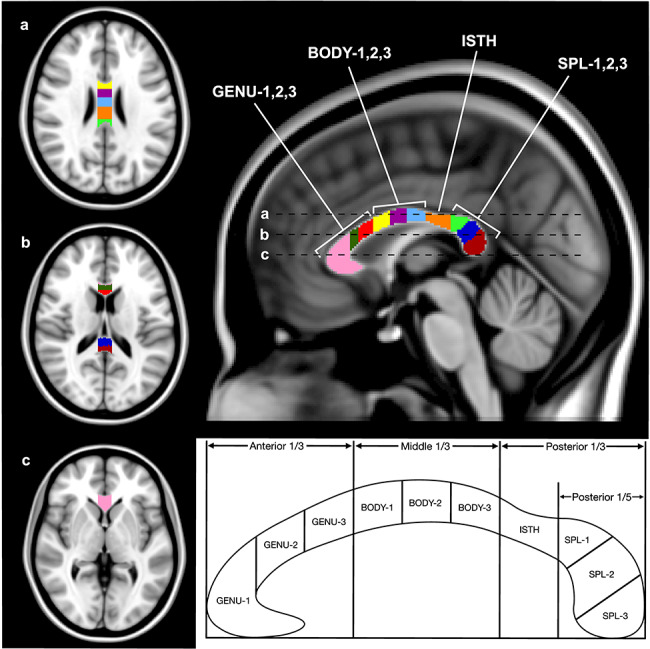
Figure 1.

Axial and midsagittal views of the CC, with 10 ROIs shown in MNI-152 standard space. The regions, from right to left: GENU-1 (pink), GENU-2 (dark green), GENU-3 (red), BODY-1 (yellow), BODY-2 (purple), BODY-3 (light blue), ISTH (orange), SPL-1 (light green), SPL-2 (dark blue), and SPL-3 (dark red). The three lines (a, b, and c) on the midsagittal view represent the location of the three axial slices displayed on the left. Schematic showing the division of the CC into 10 ROIs is shown below. The anterior and middle thirds of the CC were divided into three equal portions representing the three segments of the genu and body, respectively. The space between the beginning of the posterior 1/3 and the posterior 1/5 was designated as the isthmus, and the anterior 1/5 was divided into three equal segments (based on straight length) for the three segments of the splenium.
