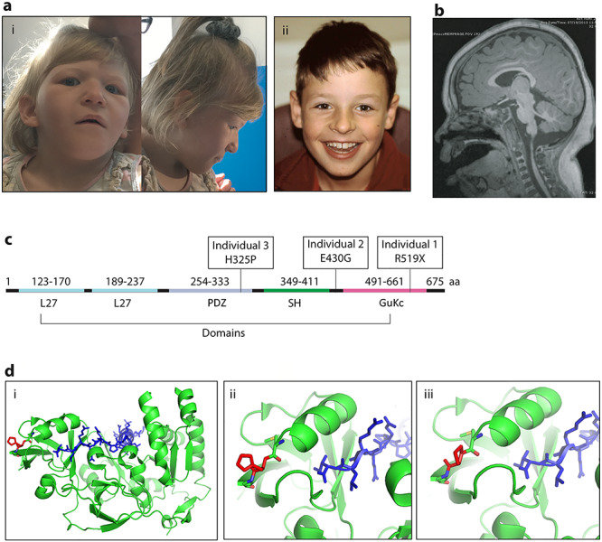
Figure 1.

Patient clinical phenotypes and variant localization. (A) Images of dysmorphic features seen in Individual 1 (i) and Individual 2 (ii). (B) Brain MRI image of Individual 1, highlighting the mild decrease in gyral folding. (C) MPP5 gene with domains and patient variants noted. (D) Molecular modeling of the missense variant in individual 3 with (i) showing the overall protein structure of MPP5, (ii) depicting His325 and (iii) showing Pro325.
