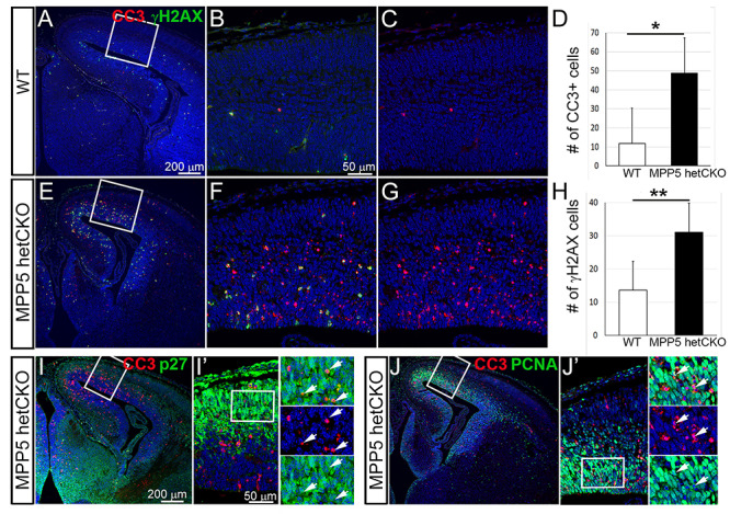
Figure 8.

Developing cerebral cortex of Mpp5 het CKO mice displays massive cell death. (A–H) Analysis of the numbers of cells undergoing apoptotic cell death labeled by CC3 (D, 313%, P = 0.12) or double-strand DNA damage marked by gH2AX (H, 128%, P = 0.001) in cortical sections of Mpp5 het CKO (E–G) and WT (A–C) at E14.5 (I–J′). Overlapping expression of CC3 and post-mitotic cell marker, p27 (I, I′) or proliferating cell marker, PCNA (J, J′) is present in Mpp5 het CKO (arrows). Data are presented as mean ± SEM. WT, n = 4; Mpp5 het CKO, n = 4.
