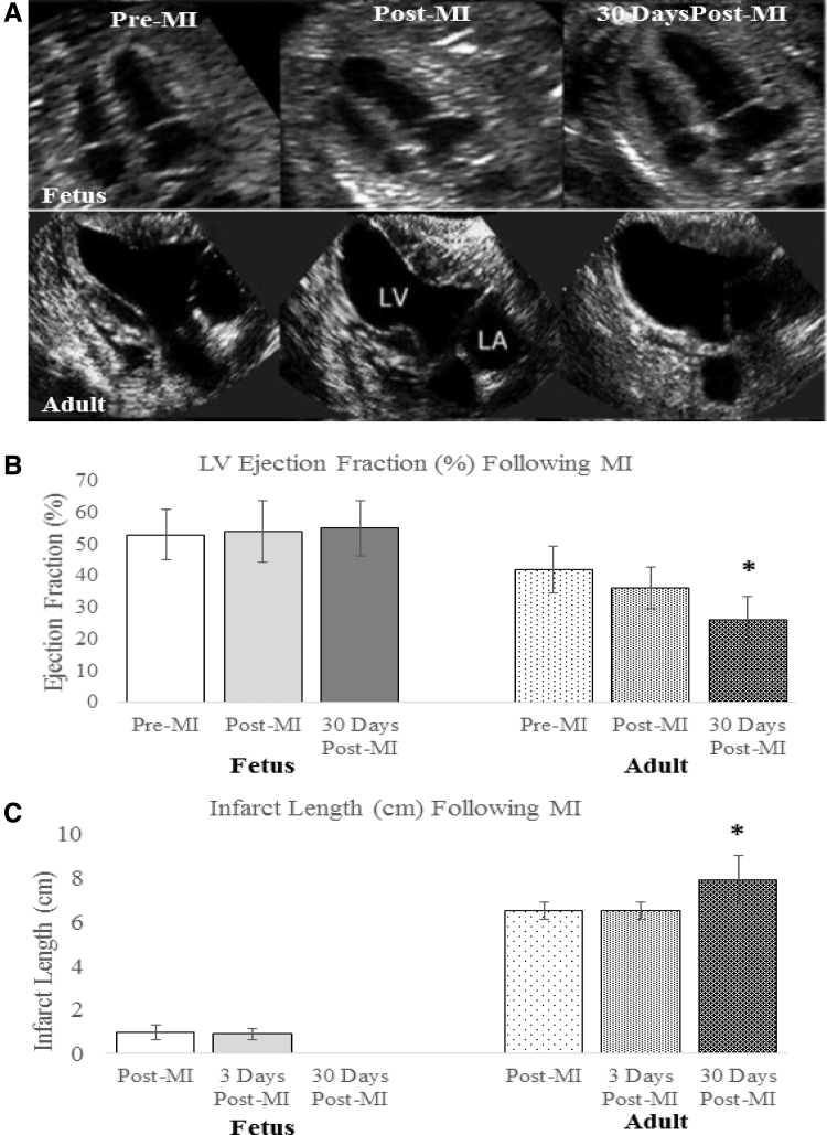
Figure 4.
Functional restoration after MI in fetal sheep (A) Cardiac function declines in the adult hearts following MI, whereas the fetal hearts show restoration of function. Serial end-systolic echocardiographic views show no evidence of LV dilation or infarcted myocardium at 30 days in the fetus, whereas the adult hearts show dilation of the LV, 30 days following infarction with a large anteroapical infarct. (B) EF measured by quantitative echocardiography is unchanged in the fetus at 3 days (p = 0.37) and 30 days (p = 0.31) following infarction. In the adult, the EF has significantly declined by 30 days following MI (*p < 0.05 vs. adult pre-MI and post-MI). (C) Absolute infarct length defined as the length of akinetic myocardium measured by echocardiography is unchanged at 3 days following infarction (p = 0.72), but decreases to zero in the fetus at 30 days following infarction (*p < 0.05 vs. fetal post-MI). In the adult, the absolute infarct length is also unchanged at 3 days following infarction (p = 1.00), but increases over a period of 30 days following infarction (*p < 0.05 vs. adult post-MI). EF, ejection fraction; LV, left ventricular.

