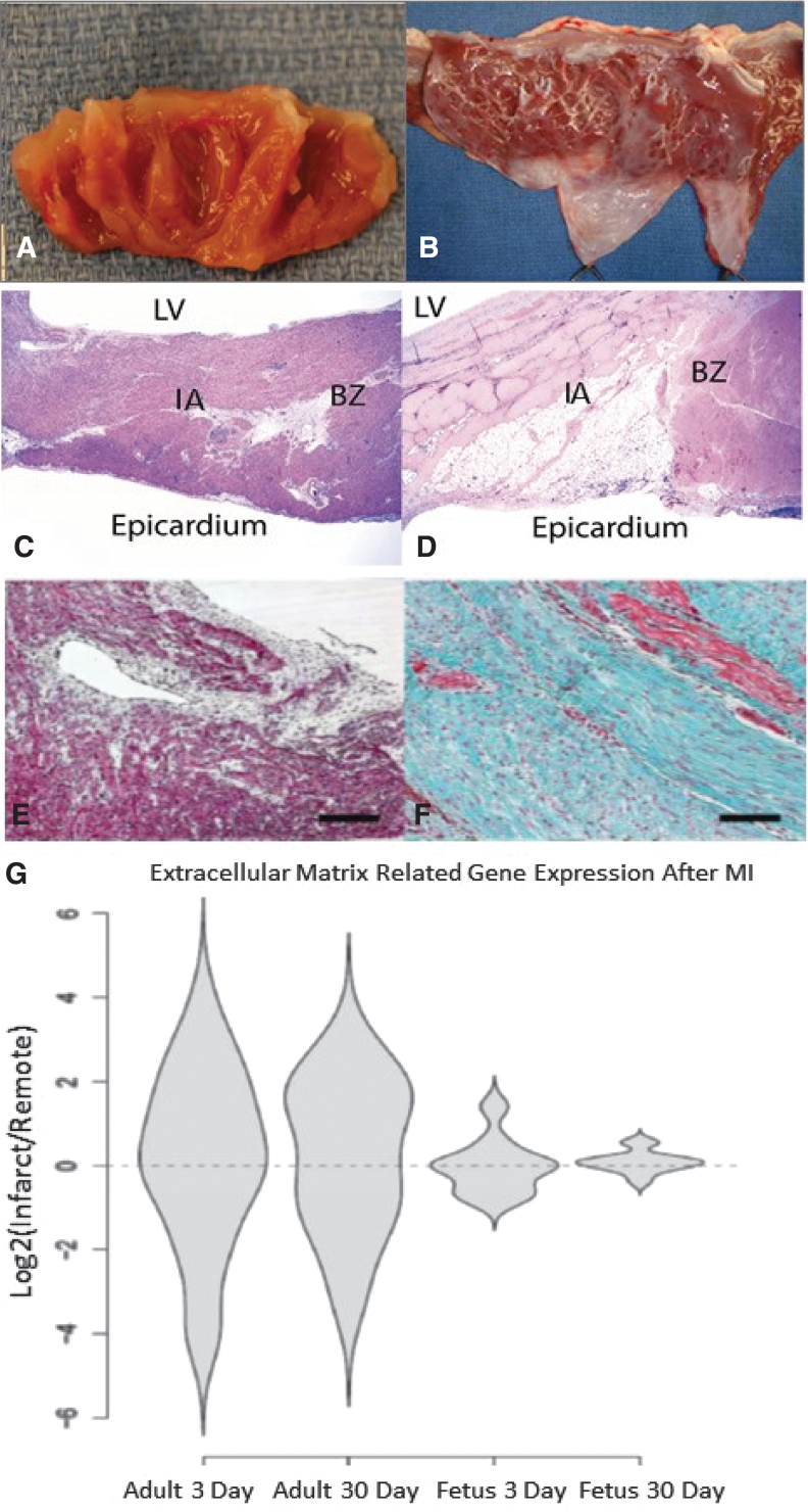
Figure 5.
Differential regulation of ECM after MI (A–F) Fetal hearts regenerate without scar formation following MI. Four weeks after myocardial infarction, (A) fetal hearts show no gross evidence of fibrosis, while (B) adult hearts show apical fibrosis and ventricular wall thinning. H&E staining at 4 weeks demonstrates (C) no evidence of myocyte loss or ventricular wall thinning in the fetal heart (I) infarct or BZ and (D) significant myocyte loss and ventricular wall thinning in the adult infarct (I) (20 × ). Masson's trichrome staining at 4 weeks following MI confirms that there is (E) minimal fibrosis in the fetal infarct (100 × ) and (F) an exuberant fibrotic response in the adult infarct (100 × ). (G) Adult infarcts demonstrated a persistent increase in the expression of “extracellular matrix” genes from day 3 to day 30, whereas the fetal infarct gene expression returned to baseline by 30 days. Violin plots for the genes related to the gene ontology term ECM. The y-axis represents the log2 of the ratio of the infarct to remote region average gene expression. The violin shapes represent the distribution of the log2 ratios in each group. (20 genes; p < 0.005, Student's t test).

