Figure 6.
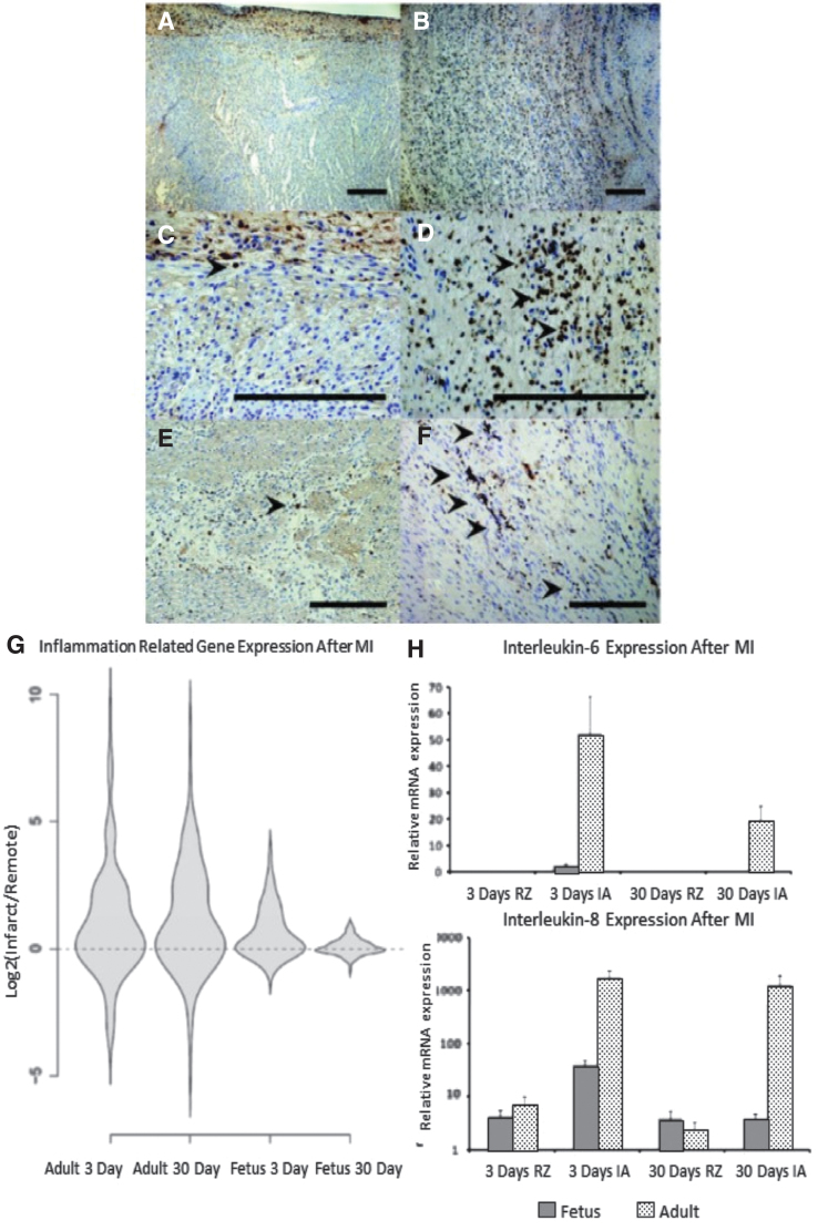
Differential inflammatory response after MI (A–F) Decreased infiltration of inflammatory cells following myocardial infarction in fetal verses adult hearts. Seven days following infarction, CD45 immunohistochemistry demonstrated that the fetal heart (A-100 × , C-400 × ) shows minimal numbers of inflammatory cells, while the adult heart (B-100 × , D-400 × ) shows a large infiltrate, with CD45 positive cells marked by arrowheads. At 4 weeks following infarction, the number of inflammatory cells in both the (E) fetal and (F) adult hearts has decreased, but the adult heart has persistent scattered areas of inflammation not seen in the fetus (200 × ). (G) Adult infarcts demonstrated a persistent increase in the expression of “inflammatory response” genes from day 3 to day 30, whereas the fetal infarct gene expression returned to baseline by 30 days. Violin plots for the genes related to the GO term “inflammatory response.” The y-axis represents the log2 of the ratio of the infarct to remote region average gene expression. The violin shapes represent the distribution of the log2 ratios in each group. (162 genes; p < 0.005, Student's t test). (H) High expression of proinflammatory cytokines IL-6 and IL-8 in adult infarcts versus fetal infarcts. Real-time PCR analysis of mRNA for IL-6 and IL-8 in fetal and adult hearts at 3 and 30 days after MI. Both IL-6 and IL-8 are highly expressed in the adult infarct 3 and 30 days after MI. However, in the fetus, 3 days after MI, IL-6 gene expression is slightly increased and completely disappears after 30 days.

