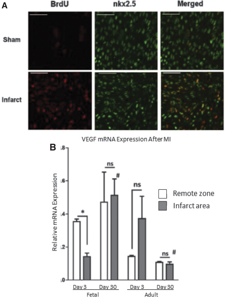
Figure 7.
Differential cellular response after MI (A) Cardiac cell proliferation contributes to fetal cardiac regeneration following myocardial infarction (MI). Representative immunohistochemistry images for 5-bromo-2-deoxyuridine (BrdU; red) and nkx2.5 (green) 3 days after MI of the borderzone region of sham and fetal infarcts. (B) VEGF-α is upregulated in fetal infarcts. Expression of VEGF-α in the fetal and adult hearts after MI. Real-time PCR analysis of VEGF-α expression in the IA (shaded bars) and RZ (unshaded bars) of fetal heart (day 3, n = 5; day 30, n = 5) and adult heart (day 3, n = 4; day 30, n = 4) at 3 and 30 days after myocardial infarction. The VEGF-α gene expression was normalized to 18S gene expression. Two-way analysis of variance followed by Fisher's least significant difference post hoc test was used to analyze the data. *p < 0.05 comparing RZ to IA in each group. #p < 0.0001 comparing RZ on day 3 to RZ on day 30 or IA on day 3 to IA on day 30. (D = day; mRNA = messenger ribonucleic acid; ns = not significant.) VEGF, vascular endothelial growth factor.

