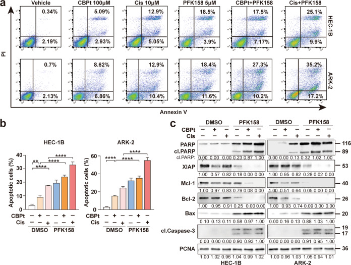
Fig. 3. Cell apoptosis induced by PFK158 and carboplatin/cisplatin in EC cells.
a HEC-1B and ARK-2 cells were treated with CBPt(100 μM)/Cis(10 μM), PFK158 (5 μM), or their combination for 24 h. The apoptotic cells were detected with Annexin-V/propidium iodide (PI) staining and analyzed by flow cytometry. Representative flow plots are shown. b Annexing-positive cells were defined as apoptotic. The data represent as mean ± SD (n = 3; **p < 0.01, ****p < 0.0001). c HEC-1B and ARK-2 cell were treated with PFK158 (5 μM), CBPt (100 μM)/Cis (10 μM) ± PFK158 for 24 h, and then the levels of PARP, cleaved caspase 3, XIAP, Mcl-1, Bcl-2 and Bax protein were determined by immunoblotting. PCNA served as a loading control.

