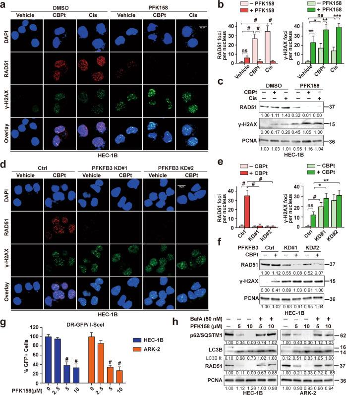
Fig. 6. PFKFB3 inhibition induces DNA damage and downregulates CBPt/Cis-induced RAD51 to disrupt DNA repair in EC cells.
a Confocal analysis of RAD51 foci (red) and γ-H2AX foci (green) in HEC-1B cells, following treatment with CBPt (100 µM)/Cis (10 µM), PFK158 (5 µM) or their combination for 24 h. n = 3 independent experiments. Scale bar, 10 μm. b Bar chart showing RAD51 foci (left panel) and γ-H2AX foci (right panel) as quantified using CellProfiler, n > 100 cells/treatment. The data represent as mean ± SD of three independent experiments, *p < 0.05; **p < 0.01; ***p < 0.001; #p < 0.0001; ns not significant. c Western blotting was performed to determine the expression of RAD51 and γ-H2AX proteins in treated HEC-1B cells. PCNA was used as a loading control. These experiments were also done in HEC-1B cells after treatment with or without CBPt (100 µM) for 24 h after PFKFB3 KD (d–f). g I-SceI expression plasmid was transfected into HEC-1B and ARK-2 cells carrying DR-GFP, and the fraction of GFP positive cells after treatment with PFK158 (0, 2.5, 5, 10 μM) for 24 h were measured by flow cytometry, with untreated cells as control (100%). The data represent as mean ± SD (n = 3, #p < 0.0001). h Western blot analysis of autophagy-related proteins (p62/SQSTM1 and LC3B) and RAD51 in HEC-1B and ARK-2 cells treated with PFK158 (5, 10 μM) for 24 h in the presence or absence of bafilomycin A (50 nM) treatment for the last 12 h. PCNA was used as the loading control.

