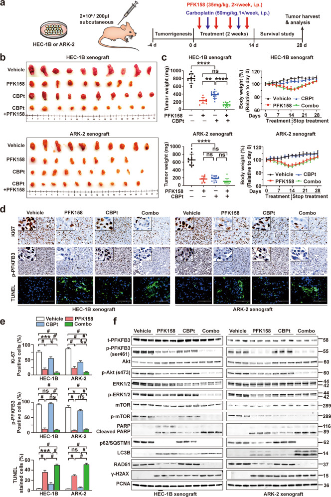
Fig. 7. Antitumor efficacy of PFK158 alone and in combination with carboplatin in two mouse xenograft models.
a Schematic diagram displaying the time course of tumor induction and treatment in mice. b Representative xenograft tumor images from each group are shown. c Effect of single-agent and dual treatment of PFK158 and CBPt on tumor growth of HEC-1B and ARK-2 cells in nude mice (n = 8–10 per group). Final tumor weights from different groups at the time of sacrifice are shown (left panel). The changes in the mouse body weights during the experiment were recorded (right panel). d IHC analysis for evaluating the expression of Ki67 and p-PFKFB3 in the tumors from vehicle or treated mice. Tumor tissues were also stained with TUNEL (green) and DAPI (blue) to investigate cell apoptosis. Scale bar, 100 μm. e Quantitative analyses of Ki67, p-PFKFB3, TUNEL staining in mouse xenograft tissues across treatment groups. Each bar represents the mean ± SD of three independent experiments, *p < 0.05, **p < 0.01, ***p < 0.001 and #p < 0.0001 for the indicated comparison; ns not significant. f Western blot analysis was performed to assess t-PFKFB3, p-PFKFB3, Akt, p-Akt, ERK1/2, p-ERK1/2, mTOR, p-mTOR, PARP, p62, LC3B, RAD51 and γ-H2AX expression in lysates from the xenografts in each group with PCNA as a loading control. Western blot was carried out in triplicate per treatment.

