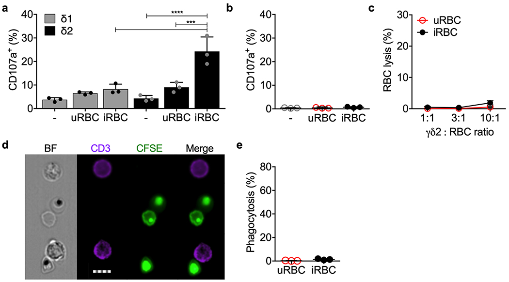
Extended Data 5. γδ1 T cells and freshly isolated healthy donor peripheral blood γδ2 T cells do not respond to iRBCs.

a, Vδ1 and Vδ2 T cells, enriched by positive selection from 3 HD and cultured for 5 days in medium containing IL-2 and IL-15, were co-cultured with uRBCs or iRBCs or no added cells in the presence of anti-CD107a. Cell degranulation was measured by CD107a staining. b-e, Highly purified freshly isolated HD γδ2 T cells from 3 donors were added to uRBCs or iRBCs to assess degranulation by CD107a staining (b), RBC lysis (c) and phagocytosis of CFSE-labeled and Pf serum-opsonized RBC (d,e). Representative images are shown in (d) and quantification of 2 independent experiments is shown in (e). Scale bar: 7 μm (d). Statistical analysis was by one-way ANOVA (a,b), two-way ANOVA with Tukey’s multiple comparisons test (c) and two-tailed nonparametric paired t-test (e). Mean ± s.e.m. is shown. P value: ***<0.001, ****<0.0001. Data shown are representative of at least three independent experiments.
