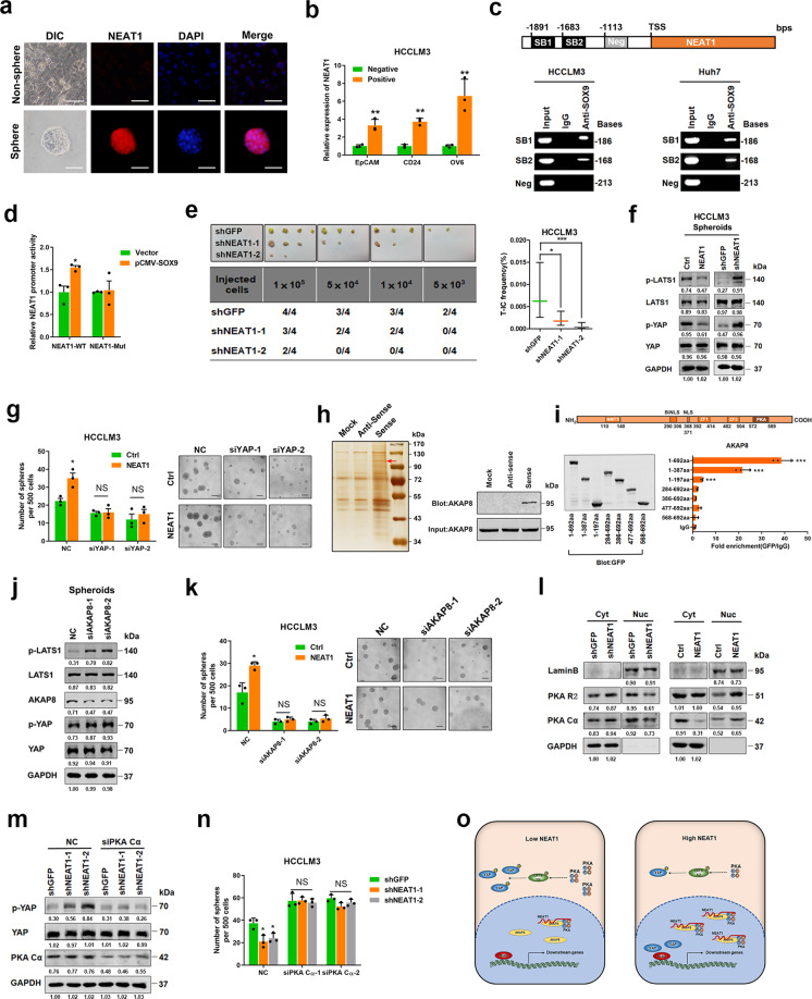
Fig. 1.
SOX9-transactived long non-coding RNA NEAT1 promotes the self-renewal of liver cancer stem cells through PKA/Hippo signaling. a The expression of NEAT1 in adherent and spheroid Huh7 cells was confirmed by FISH assays. Scale bar = 50 μm. b qRT-PCR analysis of NEAT1 in sorted EpCAM-, CD24-, or OV6-positive HCCLM3 cells relative to negative cells. c ChIP assay of the enrichment of SOX9 on NEAT1 promoter relative to IgG in HCC cells. SB indicates the SOX9-binding site and Neg indicates the region of negative control. d HCCLM3 cells were transfected with NEAT1-WT luciferase reporter plasmid or NEAT1-Mut luciferase reporter plasmid, together with pCMV-SOX9 or vector plasmid, and then subjected to luciferase reporter assay. Data were normalized against Renilla luciferase activity. e In vivo limiting dilution assay of HCCLM3 NEAT1-knockdown and control sphere-derived cells. Data are shown as the mean ± 95% CI, n = 4 for each group. f Phosphorylation of LATS1 and YAP in HCCLM3 NEAT1-overexpressing spheroids (left) and NEAT1-knockdown spheroids (right) was determined by western blot. g Sphere-formation assay of NEAT1-overexpressing cells transfected with two independent siRNAs targeting YAP. Scale bar = 100 μm. h RNA pulldown assays were performed with lysates of HCC spheres using full-length NEAT1 and antisense RNA probes, followed by mass spectrometry (left). Red arrows indicate the target band. Western blot analysis of AKAP8 in RNA pulldown precipitates retrieved by biotin-labeled NEAT1 or antisense RNA from the lysates of HCC spheres (right). i Deletion mapping for the domains of AKAP8 (upper). RIP analysis for NEAT1 enrichment in cells transiently transfected with plasmids containing the indicated EGFP-tagged full-length or truncated constructs (down). j Phosphorylation of LATS1 and YAP was determined by western blot after knockdown of AKAP8 in HCCLM3 spheroids. k The sphere-formation assay of HCCLM3 NEAT1-overexpressing cells transfected with two independent siRNAs targeting AKAP8. Scale bar = 100 μm. l Western blot analysis of PKA R2 and PKA Cα in subcellular fractions of HCCLM3 NEAT1-knockdown spheroids (left) and NEAT1-overexpressing spheroids (right). GAPDH and Lamin B acted as cytoplasm and nucleus markers, respectively. m Phosphorylation of YAP was determined by western blot after knockdown of PKA Cα in HCCLM3 NEAT1-knockdown cells. n The sphere-formation assay of HCCLM3 NEAT1-knockdown cells transfected with two independent siRNAs targeting PKA Cα. Scale bar = 100 μm. o The schematic model of the mechanism underlying the role of NEAT1 in LCSC expansion. *P < 0.05, **P < 0.01, and ***P < 0.001

