Figure 2.
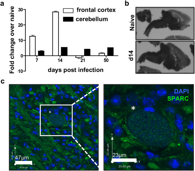
Expression of SPARC in the CNS during chronic T. gondii infection. (a–c) C57Bl/6 mice were infected with the Me49 strain of T. gondii and sacrificed at various timepoints. (a) Frontal cortex and cerebellum of infected brains were removed at days 7, 14, 21, and 50 post-infection. RNA was isolated, reverse transcribed and analyzed for levels of SPARC transcripts using qRT-PCR. Results are shown as fold change over naïve normalized to HPRT. n = 3 mice per group. Data are represented as mean ± SEM. (b) Brains were removed from naïve and 14 day infected mice. In-situ hybridization was performed on brain slices with probes specific to SPARC mRNA. (c) Confocal fluorescence microscopy of 20 μm brain slices
taken from mice at 3 weeks post infection. Image and inset show a parasite cyst (*) identified by punctate DAPI (blue) stain surrounded by parenchymal SPARC (green).

