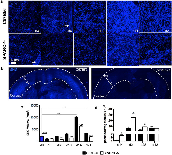
Figure 3.
SPARC−/− mice have compromised reticular fiber network in the brain during chronic T. gondii infection. C57Bl/6 and SPARC−/− mice were infected and sacrificed at 3, 6, 10, 14, and 21 days following infection. Brains were removed and imaged on a multiphoton microscope for second harmonic generation (SHG) signal. (a) 30 μm z-stacks were obtained at various locations of the cortex. 3D images were compiled and reticular fibers were analyzed for volume (c). Blood vessels (white arrows) were excluded from analysis. (b) Brains were excised from mice at 14 days post infection and images of whole coronal sections were generated by stitching overlapping Z stacks collected over the entire section. (d) C57Bl/6 and SPARC−/− mice were infected and sacrificed at 14, 21, 28, and 42 days following infection. DNA was isolated from the brain and analyzed for parasite burden using qPCR. Results are displayed as parasites per mg tissue. Data are representative of at least 2 individual experiments with a minimum of n = 4 mice per group and are represented as mean ± SEM. Volumes and parasite burden were analyzed using the student’s T test, ***p < 0.001.

