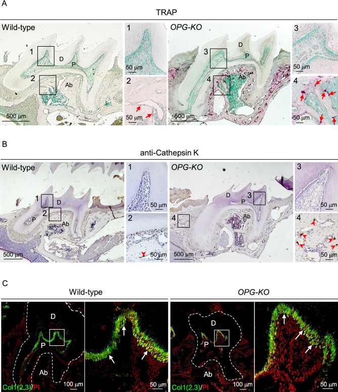
Figure 2.
OPG is not required to maintain the pulp environment in normal tooth. (A, B) Representative images of 6-week-old mouse maxillary first molars from wild-type and OPG-KO mice stained with TRAP (A) and anti-cathepsin K antibody (B). The tissues were counter stained with methyl green (A) or hematoxylin (B). Red arrows: TRAP+ osteoclasts, red arrowheads: cathepsin K+ osteoclasts. Numbered panels are magnified views of boxed areas. n = 3. (C) Representative confocal images of maxillary first molars from 6-week-old Col1(2.3)-GFP; wild-type and Col1(2.3)-GFP; OPG-KO mice. Nuclei were visualized using PI. White arrows: Col1(2.3)-GFP +odontoblasts. Right panels are magnified views of boxed areas. n = 3. P: pulp, D: dentin, Ab: alveolar bone.

