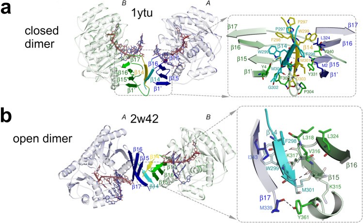
Figure 1.
Dimerization of AfAgo. (a,b) Protein subunits are coloured blue (protein chain A) and green (protein chain B). The interface-forming secondary structure elements are highlighted and numbered according to the PDB ID 2w42 assignment made by PDBsum52. The “guide” DNA/RNA strands bound by AfAgo are coloured red, “target” strands—blue. Residues 296–303 deleted in AfAgoΔ are coloured cyan and yellow. Hydrogen bonds are shown as dashed lines. (a) AfAgo complex with dsRNA (PDB ID 1ytu, both protein chains as present in the asymmetric unit), the “closed” dimer19. (b), AfAgo complex with dsDNA (PDB ID 2w42)21)—the “open” dimer. β-strands from both subunits assemble into a closed β-barrel structure, with intersubunit interface formed by β14 and β15 strands of neighbouring subunits.

