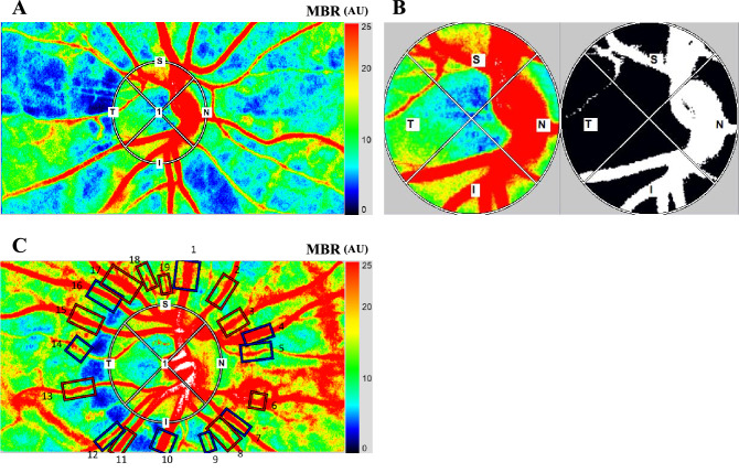
Figure 1.
A circular marker was set surrounding the optic nerve head (ONH) to measure the mean blur rate (MBR) (A). The “vessel extraction’’ function of the software then identified the vessel and tissue areas on the ONH so that the MBR could be assessed separately as the vessel areas (MBR-vessel) and the tissue areas (MBR-tissue) (B). The rectangular areas (1 to 19) were set semi-automatically to the vessels around the optic nerve head concentrically and the total retinal flow index (TRFI) was calculated (C).

