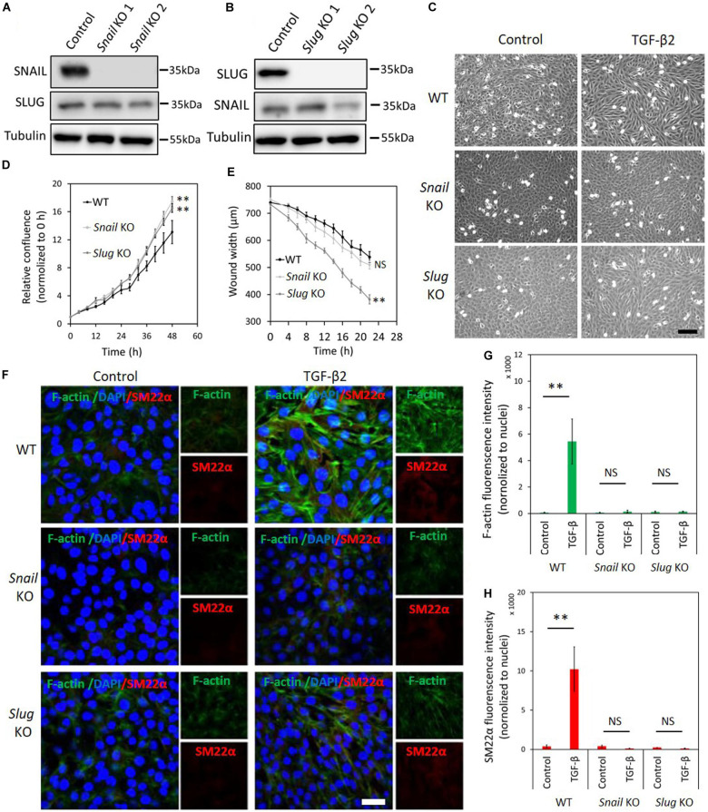
FIGURE 4.
Depletion of Snail or Slug attenuates TGF-β2-induced EndMT in 2H11 cells. (A,B) Western blot analysis of the SNAIL (A) and SLUG (B) depletion with two independent guide RNAs using CRISPR/Cas9-based gene editing in 2H11 cells. (C) Assessing cell morphological changes induced by TGF-β2 in parental 2H11, Snail knockout 2H11 and Slug knockout 2H11 cells. Brightfield microscopy images of parental 2H11 (upper panel), Snail knockout 2H11 (middle panel) and Slug knockout 2H11 (lower panel) cells showing distinct cell morphologies (i.e., cobblestone or fibroblast-like) after TGF-β2 (0.2 ng ml–1) treatment for 3 days. Scale bar: 200 μm. (D) The effects of Snail and Slug depletion on 2H11 cells proliferation. (E) The effects of Snail and Slug depletion on 2H11 cells migration ability. **p < 0.005. (F) Fluorescence microscopy analysis of mesenchymal markers in 2H11 cells. Parental 2H11, Snail knockout and Slug knockout 2H11 cells were incubated in medium containing TGF-β2 (1 ng ml–1) or medium containing ligand buffer (control) for 3 days. The expression of mesenchymal cell markers F-actin (green) and SM22α (red) in nuclei (blue) stained parental 2H11 (upper panel), Snail knockout 2H11 (middle panel) and Slug knockout 2H11 (lower panel) cells were assessed by using immunofluorescent staining. Scale bar: 50 μm. (G,H) Quantified mean fluorescence intensity of F-actin (G) and SM22α (H). At least six representative images from three independent experiments were quantified. Results are expressed as mean ± SD. NS, not significant; **p < 0.005.

