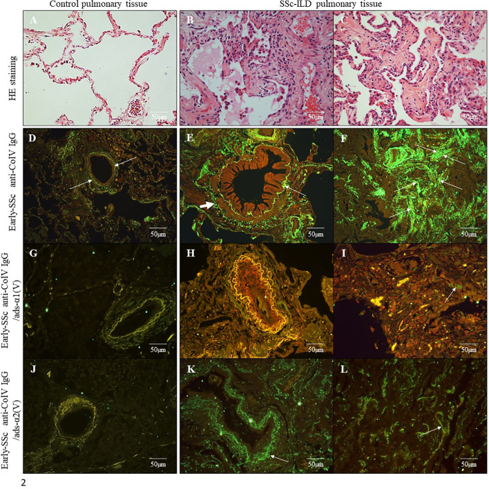
Figure 2.
Histological sections of lung samples from control subjects without associated pulmonary pathology (A) and from SSc-ILD patients with a histological pattern of Non-Specific Interstitial Pneumonia (NSIP) (B, C) (Hematoxylin & Eosin staining; original magnification: A–C, X400). Immunofluorescence in lung sections from control samples (D, G, J) and SSc-ILD patients (E, F, H, I, K, L) immunostained with early-SSc biotinylated anti-ColV IgG (D–F), anti-ColV IgG/ads-α1(V) (G–I) and anti-ColV IgG/ads-α2(V) (J–L). Note the green immunofluorescence along of periadventitial layer of the bronchovascular axis (E, K) and alveolar septa (F, L) layers (arrows). The reaction was reveled with ALEXA Fluor 488 streptavidin. Original magnification: 400X, (D–L).

