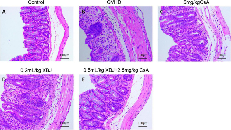
FIGURE 7.
XBJ protected intestinal tissue in aGVHD mice. Representative hematoxylin–eosin staining of colon tissues in different groups of mice was presented. (A) No-GVHD control group; (B) GVHD group; (C) 0.2 mL/kg XBJ-treated group; (D) 5 mg/kg CsA-treated group; (E) 0.5 mL/kg XBJ and 2.5 mg/kg CsA-treated group. Scale = 100 μm. n = 4–6/group.

